How We Do It: Fundamentals of Radiological Practice
How 18 - How we overcome imaging challenges in the thoracic outlet
Lectures
1
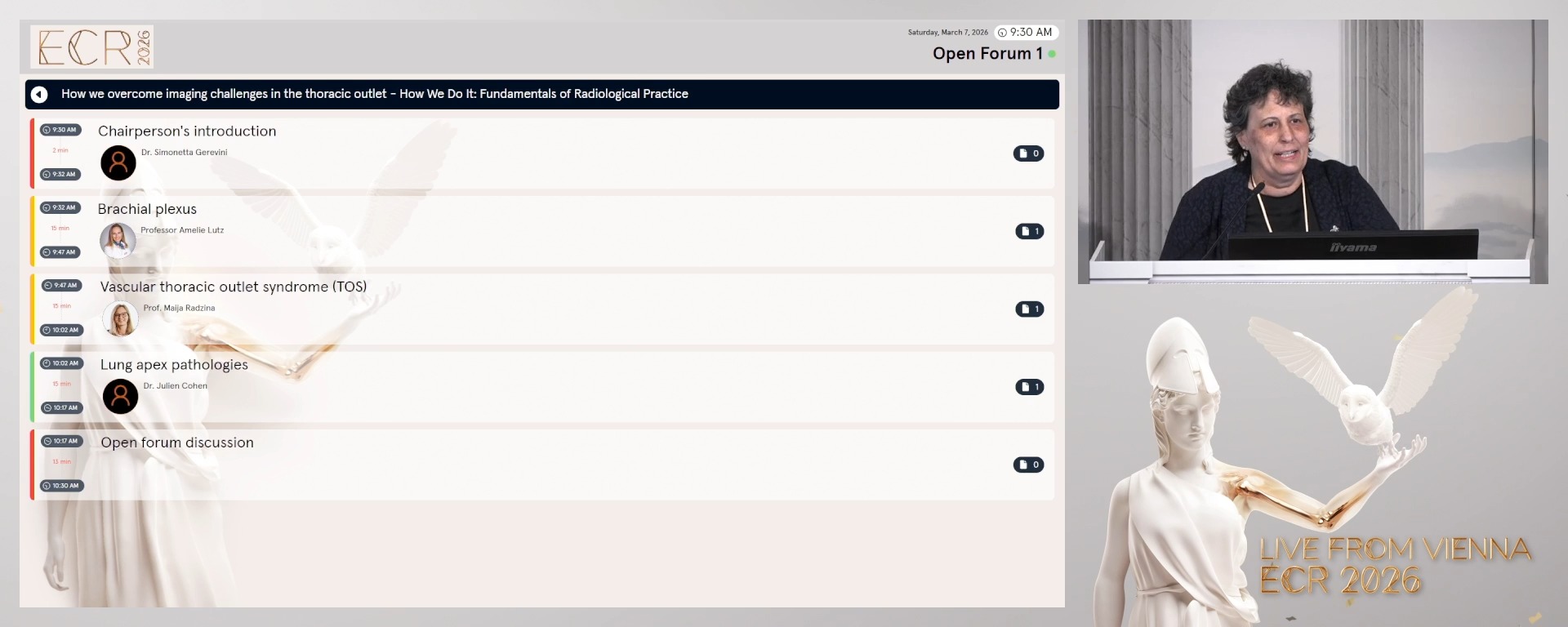
Chairperson's introduction
02:00Simonetta Gerevini, Cremona / IT
2
Brachial plexus
15:00Amelie Lutz, Kreuzlingen / CH
3
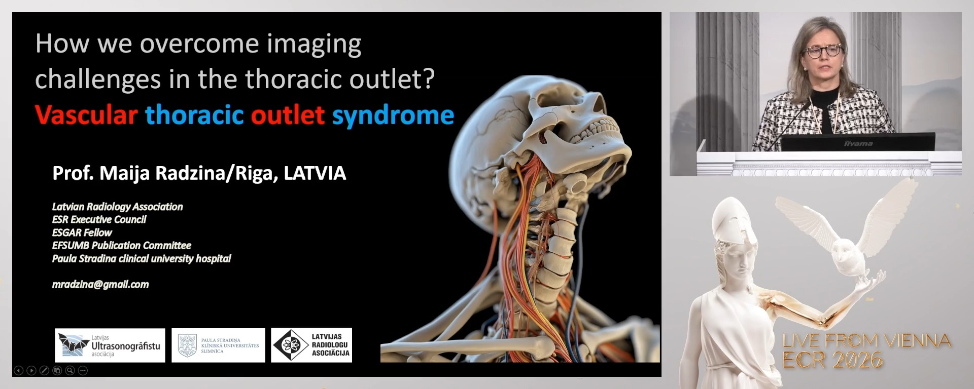
Vascular thoracic outlet syndrome (TOS)
15:00Maija Radzina, Riga / LV
4
Lung apex pathologies
15:00Julien Cohen, Geneve / CH
5
Open forum discussion
13:00Open forum discussion
2 min
Chairperson's introduction
Simonetta Gerevini, Cremona / Italy
15 min
Brachial plexus
Amelie Lutz, Kreuzlingen / Switzerland
- To learn how to tailor MRI sequences and how to mitigate artefacts for brachial plexus imaging.
- To review key anatomic landmarks on imaging and how to reliably identify the different plexus components.
- To become familiar with the most common brachial plexus pathologies and how to differentiate them from mimics using a case-based approach.
15 min
Vascular thoracic outlet syndrome (TOS)
Maija Radzina, Riga / Latvia
- To learn how to perform and interpret positional imaging studies, US, CT, and MRI for TOS evaluation.
- To understand how to choose imaging protocols tailored to neurogenic versus vascular TOS.
- To become familiar with imaging abnormalities and their clinical presentation to improve diagnostic confidence using a case-based approach.
15 min
Lung apex pathologies
Julien Cohen, Geneve / Switzerland
- To understand protocol modifications for optimised lung apex visualisation on CT and MRI scans.
- To learn about the imaging appearance of the most common benign and malignant lesions found in the lung apices.
- To understand the management strategies for incidental findings in the lung apex.
13 min
Open forum discussion