Refresher Course: Musculoskeletal
RC 610 - Imaging hip joint replacement
5 min
Chairperson's introduction
Alberto Bazzocchi, Bologna / Italy
15 min
Radiography and ultrasound: how far can you go?
Davide Orlandi, GENOVA / Italy
1. To differentiate the strengths and weaknesses of conventional radiography in assessing the structural integrity of a hip implant (THA).
2. To show the correct ultrasound examination technique of soft tissues around THA, including assessment of periprosthetic pathologic conditions such as inflammatory pseudotumor, infections, and soft tissue impingement.
3. To realise the real-time capabilities of ultrasound, providing a valuable dynamic assessment of hip muscles and tendons functional status and furnishing an excellent tool for the guidance of diagnostic and therapeutic interventional procedures, such as periprosthetic fluid collection aspiration
and postoperative hematoma drainage.
15 min
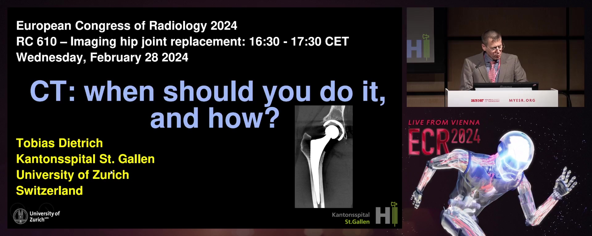
CT: when should you do it, and how?
Tobias Dietrich, St. Gallen / Switzerland
1. To present CT techniques of metal artefact reduction in hip joint replacement.
2. To show indications of CT imaging in hip joint replacement.
3. To describe hip joint replacement disorders on CT images.
2. To show indications of CT imaging in hip joint replacement.
3. To describe hip joint replacement disorders on CT images.
15 min
MRI: can it replace the above?
Amanda Isaac, London / United Kingdom
10 min
Panel discussion: Can we define an algorithm for the assessment of painful hip replacement?
