Refresher Course: Head and Neck
RC 1708 - Sinonasal and dental imaging
Lectures
1
Chairperson's introduction
05:00Soraya Robinson, Wien / AT
2
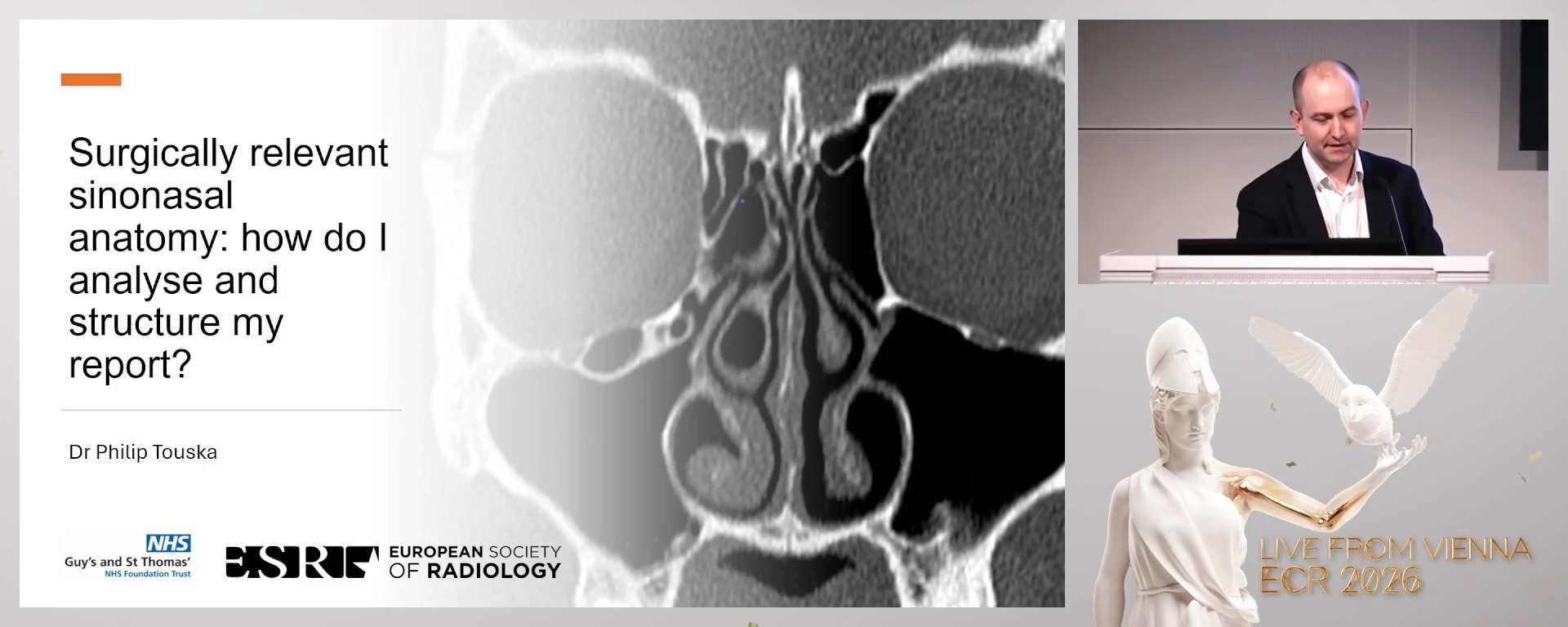
Surgically relevant sinonasal anatomy: how do I analyse and structure my report?
15:00Philip Touska, London / UK
3
Imaging complicated acute sinonasal and dental infections
15:00Jussi Hirvonen, Turku / FI
4
How I report odontogenic tumours: pearls and pitfalls
15:00Ingrid Rozylo-Kalinowska, Lublin / PL
5 min
Chairperson's introduction
Soraya Robinson, Wien / Austria
15 min
Surgically relevant sinonasal anatomy: how do I analyse and structure my report?
Philip Touska, London / United Kingdom
- To understand the complex anatomy of the sinonasal region, including critical surgical landmarks and common anatomical variants relevant to endoscopic sinus surgery.
- To learn a structured and standardised approach to analysing sinonasal CT scans using checklists or mnemonics (e.g., CLOSE) to ensure all surgically important areas are assessed and reported.
- To develop skills to produce clear, clinically relevant, and structured radiology reports that enhance communication with ENT surgeons and improve surgical planning and patient outcomes.
15 min
Imaging complicated acute sinonasal and dental infections
Jussi Hirvonen, Turku / Finland
- To recognise the imaging features of complicated acute sinonasal infections, including extension beyond the sinuses and involvement of adjacent structures on CT and MRI.
- To understand the role of imaging in differentiating sinonasal infections from dental infections and identifying potential complications such as abscess formation or osteomyelitis.
- To develop a diagnostic checklist of not-to-be-missed findings to guide timely and appropriate management of complicated sinonasal and dental infections.
15 min
How I report odontogenic tumours: pearls and pitfalls
Ingrid Rozylo-Kalinowska, Lublin / Poland
- To understand the typical imaging characteristics of odontogenic tumours (e.g., ameloblastomas, keratocystic odontogenic tumours) on CBCT, CT and MRI.
- To learn to use advanced MRI techniques, including contrast enhancement and diffusion-weighted imaging (DWI), to differentiate odontogenic tumours based on signal intensity patterns, wall thickness, and internal architecture.
- To develop a systematic approach to interpreting imaging findings to accurately distinguish between benign and potentially aggressive tumours, aiding diagnosis and treatment planning.
10 min
Panel discussion: Let’s talk anatomy: reporting that matters in the operating theatre