E³ - Young ECR Programme: Students Session
S 20 - Students Session 2
S 20-3
8 min
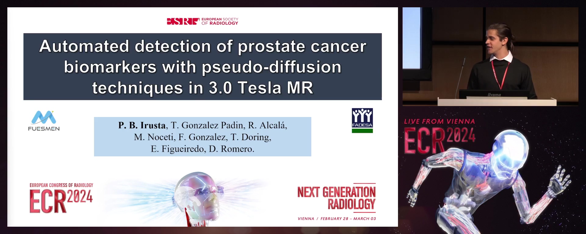
Automated detection of prostate cancer biomarkers with pseudo-diffusion techniques in 3.0 Tesla MR
Pablo Baltasar Irusta, Mendoza / Argentina
- Author Block: P. B. Irusta1, T. Gonzalez Padin1, R. N. Alcalá Marañón1, M. Noceti1, F. Gonzalez Nicolini1, T. Doring2, E. Figueiredo2, D. A. Romero3; 1Mendoza/AR, 2Rio de Janeiro/BR, 3San Carlos de Bariloche/ARPurpose: Prostate cancer (PCa) diagnosis by multiparametric MRI (mpMRI) encompasses wT2, DWI and DCE sequences. The aim of this work was to obtain significant perfusion biomarkers through IVIM-KURTOSIS (IK) with an automated pseudo-diffusion analysis achieved by post-processing DWI images employing an MR system.Methods or Background: The protocol involved 36 male subjects using a
- 0 Tesla PET/MR scanner. For the pseudo-diffusion analysis, a DWI sequence with 19 b-values (between zero and 2400 s/mm2) was implemented. Employing an in-house Python algorithm (v3.7.3), preprocessing and fitting were performed. Post-processing consisted of a three-step biexponential function fit, with automatic boundary recognition for IVIM/ADC/KURTOSIS regions and b-value thresholds for SNR. Images of DCEs (9-second temporal resolution) underwent processing using the manufacturer's workstation; IK and DCE maps were compared using the Pearson test (r>0.5). Results or Findings: Maximum b-values were constrained due to noise presence, ranging between 1900 and 2100 s/mm2 (patient dependent). The automated algorithm established region limits at 300 and 1000 s/mm
- Regarding Pearson’s coefficient, the pseudo-diffusion coefficient (D*) exhibited values of 0.88 and 0.6 for Ktrans and IAUGC, respectively, while the perfusion fraction coefficient (f) yielded 0.53 and 0.55 values. Comparing diffusion coefficient (D) and KURTOSIS (K) to IAUGC resulted in 0.58 and 0.26, respectively. Furthermore, f·D*, in comparison to Ktrans and IAUGC, showed results of 0.93 and 0.73. Conclusion: The b-value threshold for SNR was organ-proportional, as in the bladder, it decreased for the KURTOSIS region. Pearson’s coefficients indicate that IK coefficients could be considered as relevant biomarkers for the carcinoma. Obtained IK biomarkers were comparable to DCE-acquired information, given its potential inconveniences (renal dysfunction, elevated cost and vascular problems), minimising its impact.Limitations: No limitations were identified.Funding for this study: This study was performed under a research agreement with General Electric Healthcare.Has your study been approved by an ethics committee? YesEthics committee - additional information: The study was approved with the project code: 23FFUA-A
S 20-4
8 min
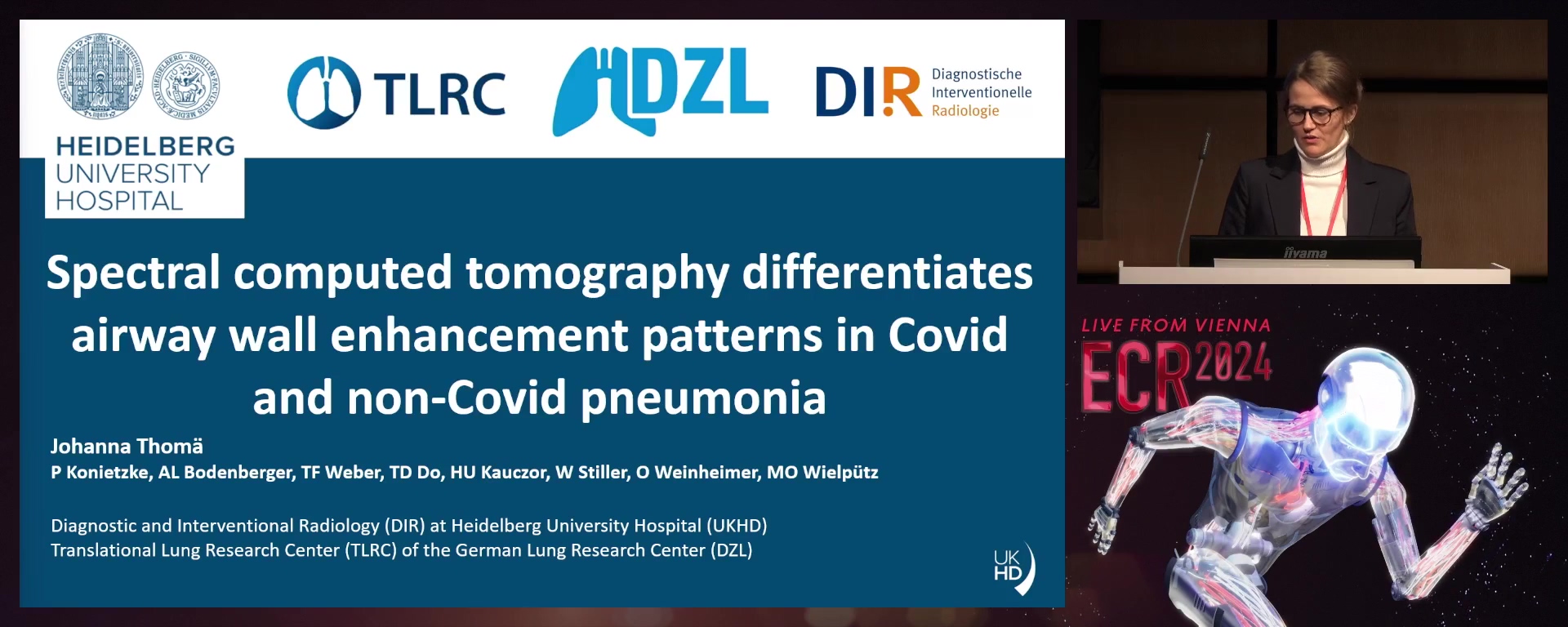
Spectral computed tomography differentiates airway wall enhancement patterns in typical and atypical pneumonia
Johanna Thomä, Heidelberg / Germany
- Author Block: J. Thomä, P. Konietzke, A. Bodenberger, T. F. Weber, T. D. Do, H-U. Kauczor, W. Stiller, O. Weinheimer, M. Wielpütz; Heidelberg/DEPurpose: Previous studies in healthy individuals demonstrated that airway wall enhancement can be quantified on virtual monochromatic reconstructions from spectral computed tomography (CT) by calculating the spectral attenuation curve’s slope based on Hounsfield Units (λHU) for wall density in segmented airways. This technique has the potential to detect airway wall inflammation in airway diseases. Thus, we aimed to differentiate patients with typical and atypical pneumonia as well as COVID-19 pneumonia from healthy individuals based on λHU.Methods or Background: 432 patients (mean age
- 9±17.2yrs) who underwent contrast-enhanced dual-layer detector spectral CT (1st and 2nd generation prototype by Philips; 303 arterial and 129 venous phase) were retrospectively recruited: 106 with typical, 107 atypical, as well as 60 with COVID-19 pneumonia, and 161 lung-healthy controls. Well-evaluated scientific software (YACTA) was used for segmenting and measuring airway wall attenuation. λHU was calculated as the median maximum airway wall attenuation at 40keV-100keV display energy and aggregated for subsegmental airway generations 5-10 (λHUG5-10). Results or Findings: λHUG5-10 was significantly higher in the arterial phase in patients with COVID-19 pneumonia (
- 2±2.3HU/keV) vs patients without pneumonia (2.1±1.2HU/keV, p<0.01), typical (2.2±1.8HU/keV, p<0.05) and atypical pneumonia (2.2±1.4HU/keV, p<0.01), but not in the venous phase. In a multivariate analysis, contrast phase (p<0.01) and pneumonia subtype were significantly correlated with λHUG5-10 (p<0.05), whereas intubation status, presence of pulmonary emboli and blood CRP were not. Conclusion: The spectral attenuation curve’s slope may differentiate airway contrast enhancement between different types of pneumonia in arterial and venous phases. It is a feasible measurement potentially representing active airway wall inflammation in pneumonia. Further studies should assess its applicability in other airway diseases such as COPD or bronchiectasis.Limitations: No limitations were identified.Funding for this study: Funding was provided by grants from the German Federal Ministry of Education and Research (82DZL00401, 82DZL004A1). Airway analysis technology is licensed to Imbio, L.L.C.. The funders and industries had no role in study design, data collection and analysis, decision to publish or manuscript preparation. The spectral CT prototype used for this study was provided by the manufacturer prior to commercial availability.Has your study been approved by an ethics committee? YesEthics committee - additional information: This retrospective study was approved by the institutional ethics committee (S-781/2018, S-924/2019, S-293/2020).
S 20-5
8 min
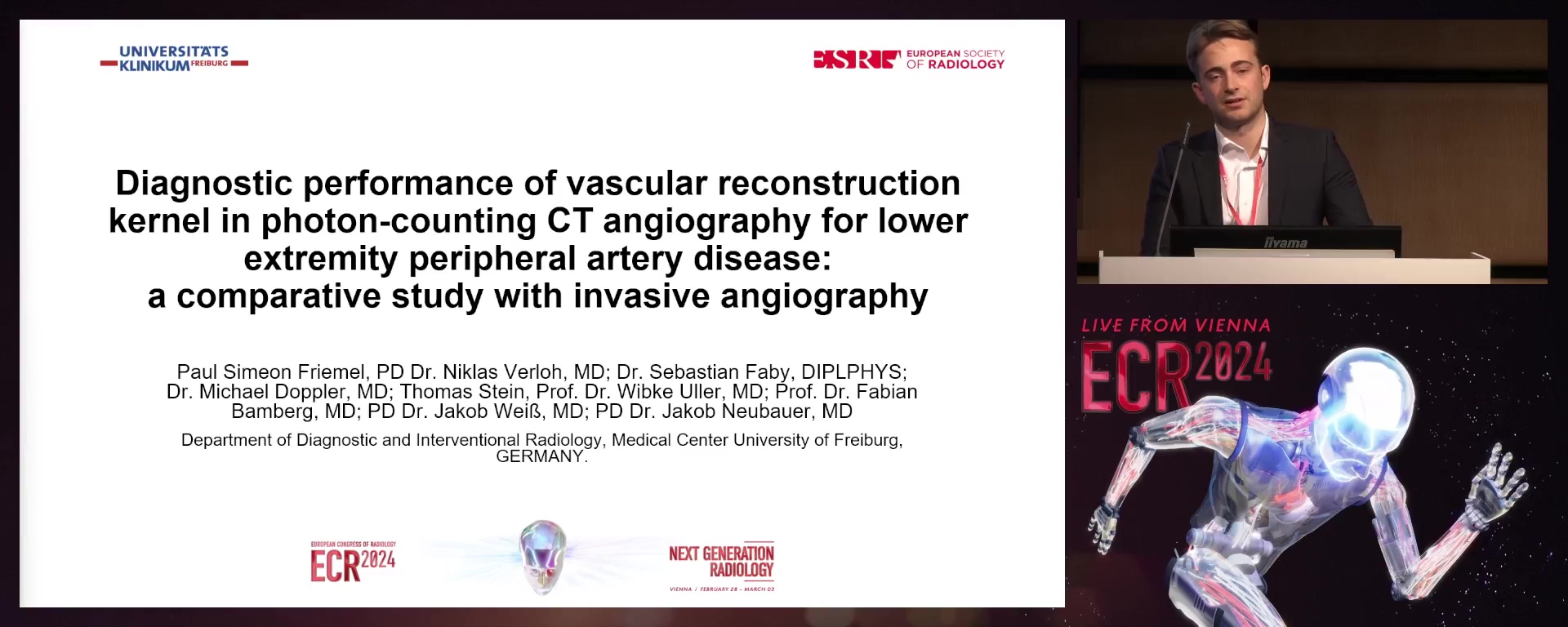
Diagnostic performance of vascular reconstruction kernel in photon-counting CT angiography for lower extremity peripheral artery disease: a comparative study with invasive angiography
Paul Simeon Friemel, Freiburg / Germany
- Author Block: P. S. Friemel, N. Verloh, J. Weiß, F. Bamberg, M. Doppler, W. Uller, S. Faby, T. Stein, J. Neubauer; Freiburg/DEPurpose: CT angiography (CTA) is vital for evaluating peripheral artery disease (PAD), but assessing lower leg vessels remains challenging due to small diameters, and impaired image quality due to calcium blooming. We aim to determine if photon-counting detector CT (PCD-CT) technology can improve the diagnosis by applying invasive digital subtraction angiography (DSA) as the reference standard.Methods or Background: In this IRB-approved study, consecutive patients with suspected lower leg PAD underwent both PCD-CT and DSA within 48 hours. PCD-CT data were reconstructed into five series using specific vascular kernels (Bv40, Bv44, Bv48, Bv56, and Bv60). DSA was performed in two orthogonal orientations. Two interventional radiologists independently assessed all PCD-CT and DSA data, randomly ordered and blinded to the reconstruction type. They rated overall image quality on a 5-point Likert scale (5=excellent) and assessed the presence and diagnostic confidence (again, 5-point Likert scale; 5=excellent) of potentially haemodynamically relevant stenosis (≥50%).Results or Findings: In the final analysis of 24 patients (age 70±11, 39% female), six ≥50% potentially haemodynamically relevant stenoses were detected using DSA. The Bv56 and Bv60 kernels provided the best overall image quality (4 [4-4]; 4 [3-5]; p≤
- 001), followed by softer kernels. The Bv56 kernel yielded the highest sensitivity (83.33%) and specificity (94.12%) for detecting potentially relevant stenosis with the highest diagnostic confidence (4 [3-5]; p≤0.001) and inter-reader agreement (k=0.7), followed by Bv60 and softer kernels. Conclusion: PCD-CT CTA with a sharp vascular kernel (Bv56) effectively detects lower leg vasculature stenosis, offering high diagnostic accuracy and confidence. These results may enhance CTA's role in evaluating patients with (suspected) PAD, potentially reducing the need for invasive DSA.Limitations: The limitations for CTA are due to small vessel calibre and potential blooming artifacts.Funding for this study: No funding was received for this study.Has your study been approved by an ethics committee? YesEthics committee - additional information: The study was approved by the ethics committee application number: 21-
S 20-6
8 min
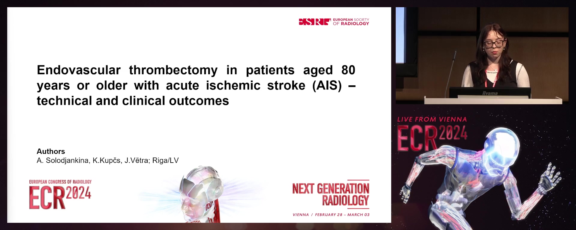
Endovascular thrombectomy in patients aged 80 years and older with acute ischaemic stroke (AIS): technical and clinical outcomes
Anastasija Solodjankina, Riga / Latvia
- Author Block: A. Solodjankina, K. Kupcs, J. Vetra; Riga/LVPurpose: This study aims to investigate the technical and clinical outcomes of endovascular thrombectomy using the thrombolysis in cerebral infarction (TICI) scale, Modified Rankin Scale (mRS) and NIH stroke scale scores in patients over 80 years with acute ischaemic stroke compared to patients in the age group under 80 years.Methods or Background: A total of 318 patients with AIS who underwent endovascular thrombectomy in Pauls Stradiņš Clinical University Hospital during the period from 2020 to 2021 were included in this retrospective cohort study. All patients were divided into two groups based on their age: those aged under 80 (235) and those aged 80 and above (83). In this study, the statistical analysis of clinical and technical outcomes of both groups measured by three scales (HIHSS, mRS, TICI) was performed using IBM SPSS.Results or Findings: There was a significant difference between the two groups in terms of pre- and postoperative NIHSS values (p=
- 045) and no significant difference between the two groups in terms of pre- and postoperative mRS scores (p=0.113). The TICI score was not significantly different between the two groups (p=0.241). The mortality rate after endovascular thrombectomy is higher in elderly patients (>80 years), 19.28%, compared to younger patients (<80 years), 11.91%. Conclusion: The findings of this study show that the impairment caused by an AIS, which is quantified with NIHSS, is more severe after endovascular thrombectomy in elderly patients (>80 years) than in younger patients (<80 years). The technical outcomes characterised by the TICI score are not significantly different between the two age groups. Disability in patients who have suffered an AIS measured by mRS is not significantly different between the two age groups.Limitations: The main limitation of this study is the multimorbidity of patients, which could cause difficulties in the interpretation of the results.Funding for this study: No funding was provided for this study.Has your study been approved by an ethics committee? YesEthics committee - additional information: The Riga Stradiņš University's ethics committee has approved this study.
S 20-7
8 min
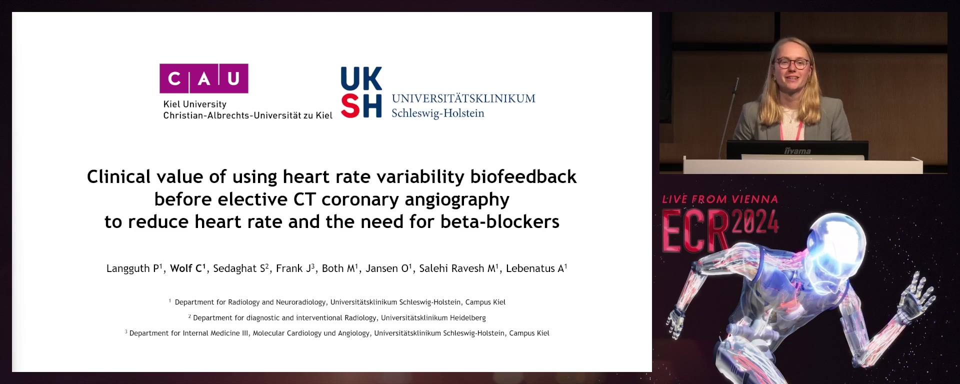
Clinical value of using heart rate variability biofeedback before elective CT coronary angiography to reduce the heart rate and the need for beta‑blockers
Carmen Wolf, Zürich / Switzerland
- Author Block: C. Wolf; Kiel/DEPurpose: Coronary computed tomography angiography (CCTA) is a commonly used method to assess coronary artery disease. Therefore, patients’ heart rate (HR) must be low and regular to minimise artifacts and radiation dose. In our prospective study, we investigated the value of heart rate variability (HRV) biofeedback before elective CCTA to reduce patients’ HR and to avoid the application of beta-blockers.Methods or Background: Sixty patients who received CCTA were included in our study and separated into two groups: with biofeedback (W-BF) and without biofeedback (WO-BF). The W-BF group used an HRV biofeedback device for 15 min before CCTA. HR was determined in each patient at four measurement time points (MTP): during the pre-examination interview (MTP1), positioning on the CT patient table before CCTA (MTP2), during CCTA image acquisition (MTP3), and after completing CCTA (MTP4). If necessary, beta-blockers were administered in both groups after MTP2 until an HR of less than 65 bpm was achieved. Two board-certified radiologists assessed the image quality and analysed the findings.Results or Findings: Overall, the need for beta-blockers was significantly lower in patients in the W-BF group than in the WO-BF group (p=
- 032). The amount of HR reduction between MTP1 and MTP2 was significantly higher in the W-BF group than in the WO-BF group (p=0.028). There was no significant difference between the W-BF and WO-BF groups regarding image quality (p=0.179). Conclusion: Application of HRV biofeedback prior to elective CCTA could decrease HR and beta-blocker use without compromising CT image quality and analysis.Limitations: Biofeedback was only performed until the patient was positioned on the CT table. We could possibly not achieve the maximal effect of HRV biofeedback during the CCTA examination itself. Integrating visual feedback into the CT scanner might further enhance and stabilise HRV biofeedback effects.Funding for this study: No funding was received for this study.Has your study been approved by an ethics committee? YesEthics committee - additional information: The study was approved by the local institutional ethics board of Kiel University (No. D 549/19).
S 20-8
8 min
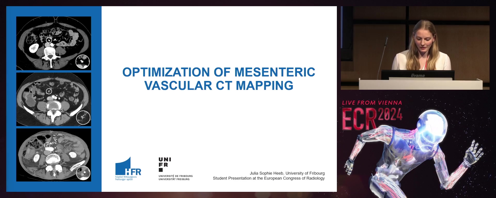
Optimisation of mesenteric vascular CT mapping
Julia Sophie Heeb, Sevelen / Switzerland
- Author Block: J. S. Heeb, H. Thoeny, L. Widmer; Fribourg/CHPurpose: The study aims to compare contrast opacification in CT angiography of mesenteric vessels between three cohorts using a fixed trigger delay, a patient-individualised split-bolus injection, and a slow contrast media injection.Methods or Background: Complete mesorectal excision in colorectal cancer is technically demanding due to frequent variations in the vascularisation of the colon. Mapping of the mesenteric anatomy could decrease surgery length and vascular lesions. We aim to optimise this preoperative mesenteric vascular mapping. In this IRB-approved single-centre prospective study, 162 consecutive patients were prospectively recruited into three cohorts with distinct CT vascular mapping protocols. The inclusion of 54 participants per cohort was determined from a-priori statistical power analyses. One reader assessed objective image quality; two readers assessed subjective image quality. The proportion of unidentified vessels was analysed using beta regression (chi-squared test). A one-way between-subject ANOVA was computed to test for differences in mesenteric vessel attenuation values.Results or Findings: Of the three protocols, the split-bolus protocol had the most extreme attenuation values. The fixed trigger delay and the slow injection protocols had more similar mean values, with the slow injection protocol showing higher attenuation values for both arteries and veins. This was expressed by significantly lower artery attenuation values in the fixed trigger delay protocol than in the other two protocols (F[2,138] =
- 7, p < 0.001) and significantly lower vein attenuation values in the split-bolus protocol than in the other two protocols (F[2,138] = 41.9, p < 0.001). The proportion of unidentified vessels was not significantly different between protocols. Conclusion: Slow and continuous contrast media injection improves opacification in CT angiography of the mesenteric vessels, providing enhanced vascular mapping for surgery.Limitations: Protocols were tested on different cohorts.Funding for this study: The study has received institutional funding.Has your study been approved by an ethics committee? YesEthics committee - additional information: The study was approved by CER-VD, swissethics.
S 20-9
8 min
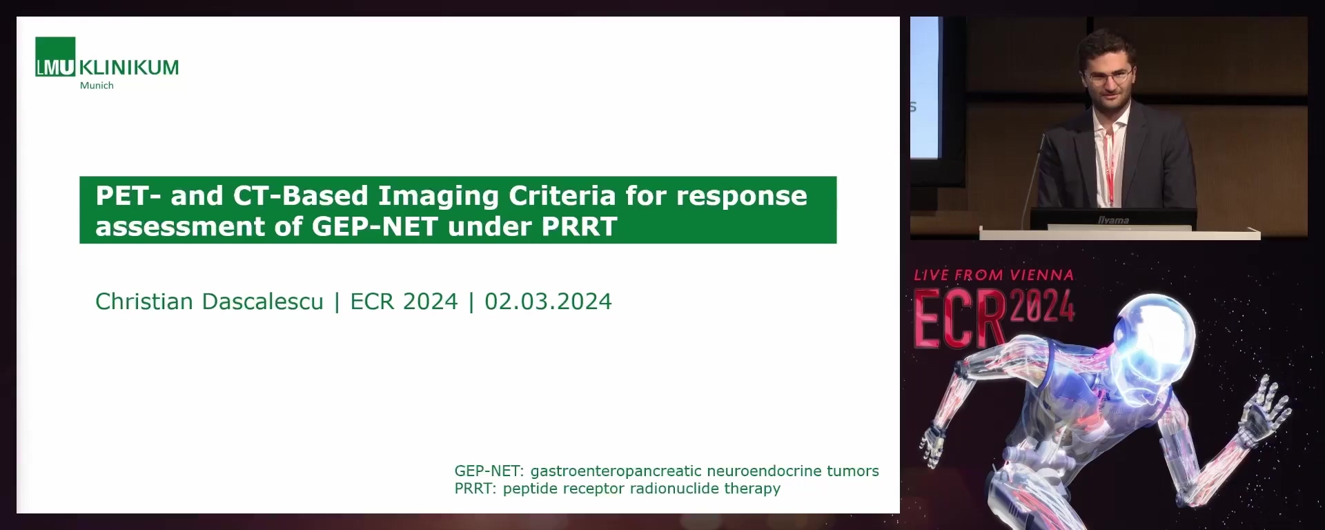
Predictive value of PET- and CT-based imaging criteria at first follow-up for GEP-NET under PRRT
Christian Alexander Dascalescu, München / Germany
- Author Block: C. A. A. Dascalescu, M. Heimer, M. P. Fabritius, L. Unterrainer, J. Rübenthaler, M. Ingrisch, J. Ricke, C. C. Cyran; Munich/DEPurpose: The study aims to evaluate and compare the predictive value of different PET- and CT-based imaging criteria on peptide receptor radionuclide therapy (PRRT) and therapy guidance in gastroenteropancreatic neuroendocrine tumours (GEP-NET) at interim follow-up.Methods or Background: In this single-centre retrospective study, we reviewed 178 patients with GEP-NET who were treated with at least two consecutive cycles of PRRT and underwent somatostatin receptor (SSR-) PET/CT at baseline and after two cycles of PRRT. We evaluated the agreement between RECIST
- 1 and mCHOI criteria to a multidisciplinary GEP-NET tumour board and assessed the modified Krenning and SSTR-RADS scores at baseline and interim follow-up. Results or Findings: The overall concordance between the response criteria was fair (weighted Cohen's Kappa=
- 26). The concordance at the follow-up between RECIST and mCHOI with the tumour-board assessment was comparable with Cohen's Kappa of 0.42 and 0.39, respectively. The survival analysis showed no significant differences in PFS for RECIST 1.1 or mCHOI categories SD and PR (log-rank p=0.70 and 0.68, respectively). mChoi-criteria showed a higher sensitivity (sensitivity: 90%, specificity: 96%, PPV: 44%, NPV: 99%) but a lower positive predictive value in predicting PRRT discontinuation due to a PD in tumour-board assessment as compared to RECIST 1.1 (sensitivity: 50%, specificity: 100%, PPV: 63%, NPV: 98%). A baseline Krenning score of 3 was associated with a shorter PFS compared to a Krenning score of 4 (721 days vs 1169 days; p=0.01). Assessment and stage migration of SSTR-RADS scores between baseline and follow-up yielded no significant value for response assessment. Conclusion: RECIST
- 1 and mCHOI have comparable diagnostic values to predict PFS. At baseline, the Krenning score could contribute to estimating long-term therapy response. Further research towards integrated bimodal monitoring and assessment tools in SSR PET/CT is warranted to guide therapy management at the interim follow-up. Limitations: The primary limitation was limited response assessment time points, including the first interim follow-up only.Funding for this study: Funding was received from the Wilhelm Vaillant Stiftung.Has your study been approved by an ethics committee? YesEthics committee - additional information: This study was approved by the ethics committee of LMU-Munich, Project number: 20-
S 20-10
8 min
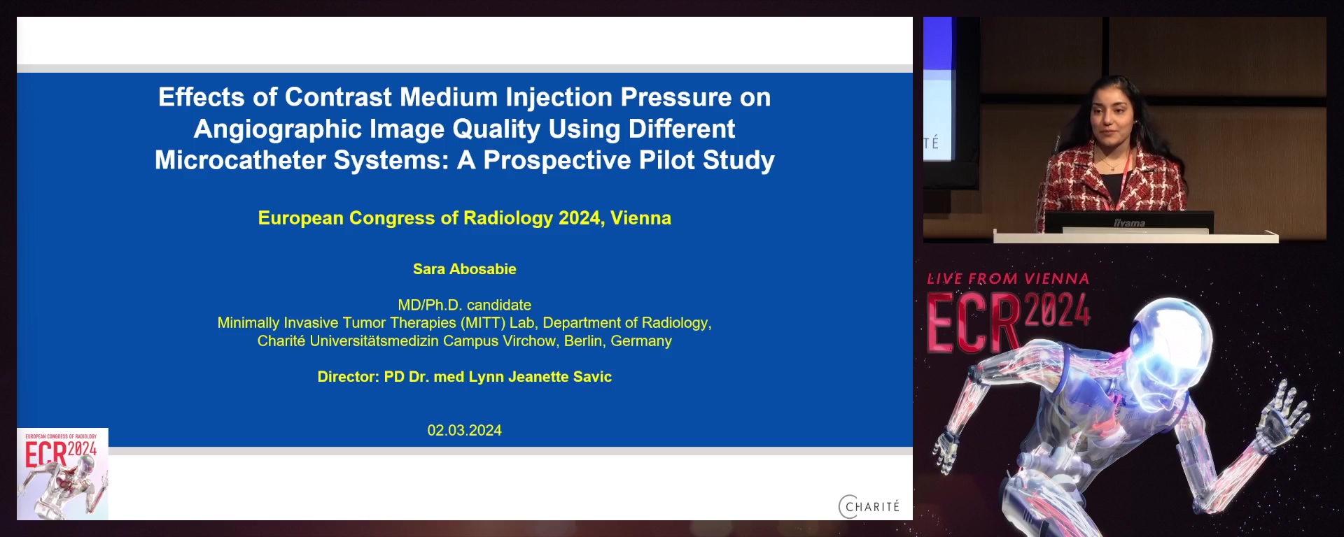
Effects of contrast medium injection pressure on angiographic image quality using different microcatheter systems: a prospective pilot study
Sara Abosabie, Berlin / Germany
- Author Block: S. Abosabie, T. Kao, D. Schnapauff, T. A. Auer, M. Jonczyk, W. Lüdemann, A. Frisch, B. Gebauer, L. J. Savic; Berlin/DEPurpose: This pilot study tested the hypothesis that a higher contrast medium (CM) injection pressure can improve the image quality of digital subtraction angiography (DSA) and reduce DSA time and radiation exposure.Methods or Background: This prospective single-centre study included 12 patients with hepatocellular carcinoma (n=11) or liver metastases (n=1) with intra-arterial therapies (10/2022-03/2023) to systematically compare DSA image quality (primary) and radiation exposure (secondary endpoint) using two microcatheters with different maximum application pressures (Pmax). IRB approval and informed consent were obtained. Patients underwent two DSAs (20ml CM, 4ml/s) using Progreat (Terumo, Pmax=750 PSI) and DraKon (Guerbet, Pmax=1200 PSI) microcatheters (both
- 4Frx130cm) placed in the common hepatic artery. Application pressure, CM flow, volume, and dose area product (DAP) were recorded. The image quality was evaluated by three blinded IR using a customised questionnaire with 16 questions in 5 categories. Responses were converted to numerical values and compared by paired t-test. Results or Findings: The mean maximum application pressure was higher using Drakon (917±94 PSI) than Progreat (731±45 PSI). In 10 patients with Progreat, the injection was terminated early. Mean DAP with DraKon (376 μGym2) was slightly lower than with Progreat (388 μGym2). Vessel visualisation was equivocal or superior with DraKon in 72% of the patients. Additionally, in 100% of patients, tumour blush was demarcated equally or more clearly in DSAs with DraKon (p<
- 001). Conclusion: Our results suggest potential benefits of standardised CM injections for DSA using higher application pressure to enhance image contrast and tumour demarcation during IAT. These findings may enable faster and more precise embolisation if confirmed in a larger cohort.Limitations: In the future, we intend to conduct a larger study with a much larger patient cohort because our cohort currently only consists of 12 patients.Funding for this study: This study was funded by a research grant from Guerbet.Has your study been approved by an ethics committee? YesEthics committee - additional information: This study was approved by the Ethics Committee of Charité Universitätsmedizin Berlin.
S 20-11
8 min
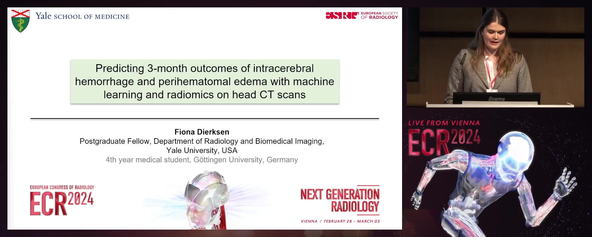
Prognostic machine learning classifiers using radiomics of supratentorial intracerebral haemorrhage and surrounding oedema on admission non-contrast head CT
Fiona Dierksen, Göttingen / Germany
- Author Block: F. Dierksen1, S. Haider1, A. T. Tran1, H. Lin1, J. Sommer1, I. Maier2, S. Aneja1, A. I. Qureshai3, S. Payabvash1; 1New Haven, CT/US, 2Göttingen/DE, 3Columbia, MO/USPurpose: Following intracerebral haemorrhage (ICH), the haemorrhagic lesion represents the extent of primary brain injury, whereas surrounding oedema represents secondary brain injury related to blood product degradation. While prior studies linked admission ICH volume and, more recently, ICH radiomic features with clinical outcomes, there have barely been studies on prognostic correlates of combined ICH and peri-haematomal oedema (PHE) radiomic features. We developed an improved algorithm for patient outcome prediction by incorporating oedema radiomics into the model.Methods or Background: Using the ATACH-2 trial dataset, we extracted 1130 features from manually segmented ICH and PHE lesions on admission non-contrast head CTs. We split the data from 892 patients into discovery (n=500) and independent validation (n=352) cohorts. In the discovery cohort, we trained, optimised, and compared the performance of 36 combinations of six machine-learning classifiers and six feature selection methodologies for the prediction of favourable vs poor outcomes based on a 3-month modified Rankin score. Separate models were trained and tested for radiomics from ICH vs ICH and PHE features. Finally, we evaluated the best-performing models using ICH vs ICH and PHE features.Results or Findings: The best-performing model using ICH features alone was with Naïve-Bayes and RIDGE regression, achieving an AUC of
- 71 (confidence interval: 0.65-0.77), and the best-performing model using ICH and oedema features was Elastic-Net and RIDGE achieving an AUC of 0.74 (CI: 0.7-0.79). Although there was no significant difference in AUCs in the validation cohort (p=0.074), in risk assessment, there was a 17% improvement in the Net Reclassification Index (p<0.001) and a 12% improvement in the Integrated Discrimination Index (p<0.001). Conclusion: Incorporating PHE in addition to ICH features from admission head CT can improve the outcome risk assessment of prognostic machine-learning models in ICH patients.Limitations: The study was limited by missing 72-hour follow-up images.Funding for this study: Dr. Payabvash is supported by the National Institutes of Health (K23NS118056) and the Doris Duke Charitable Foundation (2020097).Has your study been approved by an ethics committee? YesEthics committee - additional information: The ethics approval was ensured by the ATACH-2 investigators (ClinicalTrials.gov identifier: NCT01176565). Our group performed post-hoc analyses of anonymised data.
