Refresher Courses: Collaborative Learning - Jointly organised by the ECR Scientific Subcommittees Head and Neck and Neuro
RC 800c - The skull base: imaging essentials
Lectures
1
Chairperson's introduction
05:00Yeliz Pekçevik, İzmir / TR
2
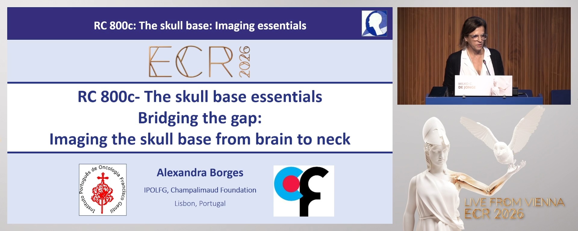
Bridging the gap: imaging the skull base from brain to neck
15:00Alexandra Borges, Lisbon / PT
3
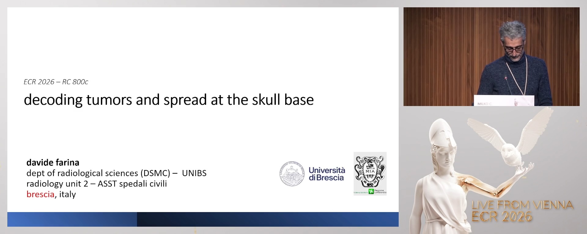
Decoding tumours and spread at the skull base
15:00Davide Farina, Brescia / IT
4
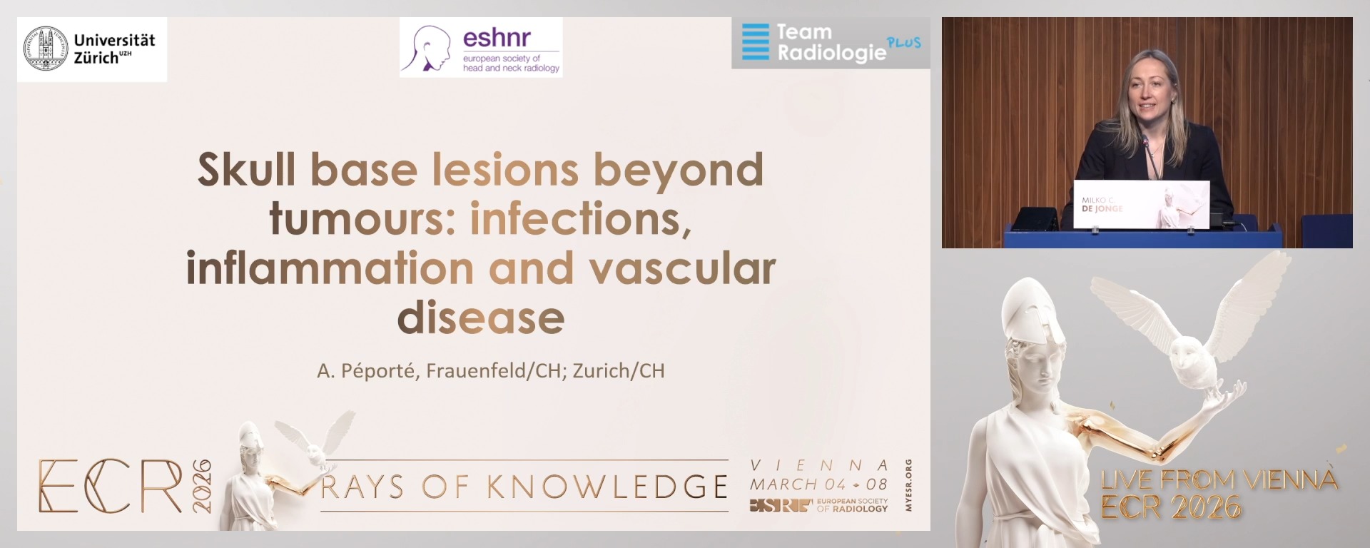
Skull base lesions beyond tumours: infections, inflammation and vascular disease
15:00Anne Renée Juliette Péporté, Frauenfeld / CH
5
Panel discussion: Future directions in skull base imaging
10:00Panel discussion: Future directions in skull base imaging
5 min
Chairperson's introduction
Yeliz Pekçevik, Izmir / Turkey
15 min
Bridging the gap: imaging the skull base from brain to neck
Alexandra Borges, Lisbon / Portugal
- To review key anatomical landmarks of the skull base relevant to both neuroradiologists and head and neck radiologists.
- To identify the most effective imaging modalities and protocols for assessing complex skull base regions.
- To understand how cross-speciality collaboration enhances diagnostic accuracy in skull base pathology.
15 min
Decoding tumours and spread at the skull base
Davide Farina, Brescia / Italy
- To differentiate common skull base tumours based on imaging characteristics and location.
- To recognise typical and atypical patterns of tumour infiltration and perineural spread.
- To apply a multimodal and multidisciplinary approach to accurately assess tumour extent and guide management.
15 min
Skull base lesions beyond tumours: infections, inflammation and vascular disease
Anne Renée Juliette Péporté, Frauenfeld / Switzerland
- To identify imaging features of infectious, inflammatory, and vascular skull base lesions.
- To distinguish between tumour-mimicking and true neoplastic processes in challenging cases.
- To understand key diagnostic pitfalls and when to recommend further imaging or biopsy.
10 min
Panel discussion: Future directions in skull base imaging