Refresher Course: Head and Neck
RC 1208 - Structured approach to skull base and temporal bone imaging
Lectures
1
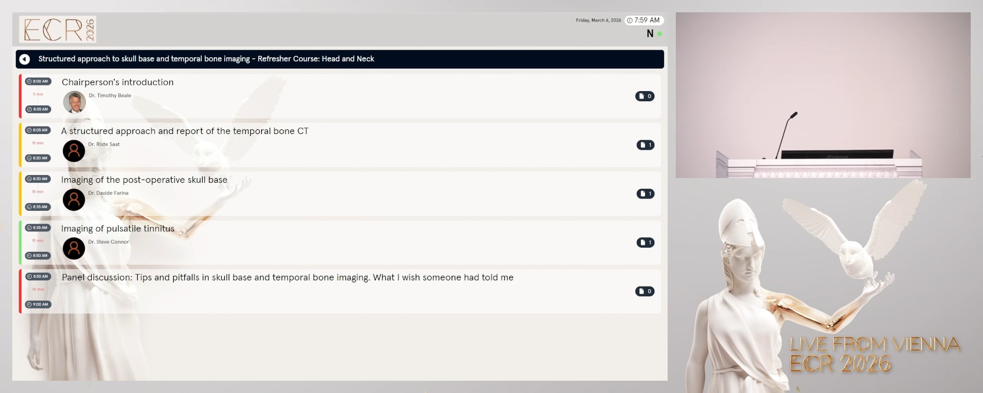
Chairperson's introduction
05:00Timothy Beale, London / UK
2
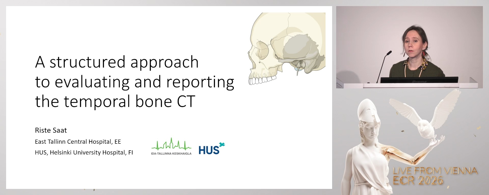
A structured approach and report of the temporal bone CT
15:00Riste Saat, Tallinn / EE
3
Imaging of the post-operative skull base
15:00Davide Farina, Brescia / IT
4
Imaging of pulsatile tinnitus
15:00Steve Connor, London / UK
5
Panel discussion: Tips and pitfalls in skull base and temporal bone imaging. What I wish someone had told me
10:00Panel discussion: Tips and pitfalls in skull base and temporal bone imaging. What I wish someone had told me
5 min
Chairperson's introduction
Timothy Beale, London / United Kingdom
15 min
A structured approach and report of the temporal bone CT
Riste Saat, Tallinn / Estonia
- To understand the complex anatomy of the temporal bone and its key structures visible on CT and learn a systematic, structured method to evaluate temporal bone CT images, starting with axial and coronal planes and including evaluation of pneumatisation, middle and inner ear structures, and critical neurovascular canals.
- To recognise common surgically relevant anatomical variants on CT imaging.
- To develop skills for clear, comprehensive CT reporting in different clinical settings.
15 min
Imaging of the post-operative skull base
Davide Farina, Brescia / Italy
- To understand the typical postoperative imaging appearances of skull base reconstructions following various surgical approaches, including open and endoscopic techniques.
- To identify and distinguish normal reconstructive tissues from recurrent or residual neoplastic disease on CT and MRI.
- To recognise common postoperative complications such as infection, haemorrhage, and graft-related changes, and understand their imaging characteristics.
- To gain practical knowledge of interpreting altered anatomy after skull base surgery to optimise patient follow-up and management.
15 min
Imaging of pulsatile tinnitus
Steve Connor, London / United Kingdom
- To understand the pathophysiology and clinical significance of pulsatile tinnitus and its relationship to vascular and temporal bone abnormalities.
- To learn the imaging modalities and protocols best suited for evaluating patients with pulsatile tinnitus, emphasising CT and MRI techniques.
- To identify common and less common causes of pulsatile tinnitus on imaging.
- To develop a systematic approach to image interpretation to guide diagnosis and clinical management of pulsatile tinnitus.
10 min
Panel discussion: Tips and pitfalls in skull base and temporal bone imaging. What I wish someone had told me