How We Do It: Fundamentals of Radiological Practice
How 14 - How we report diagnostic and follow-up MRI scans in multiple sclerosis (MS)
Lectures
1
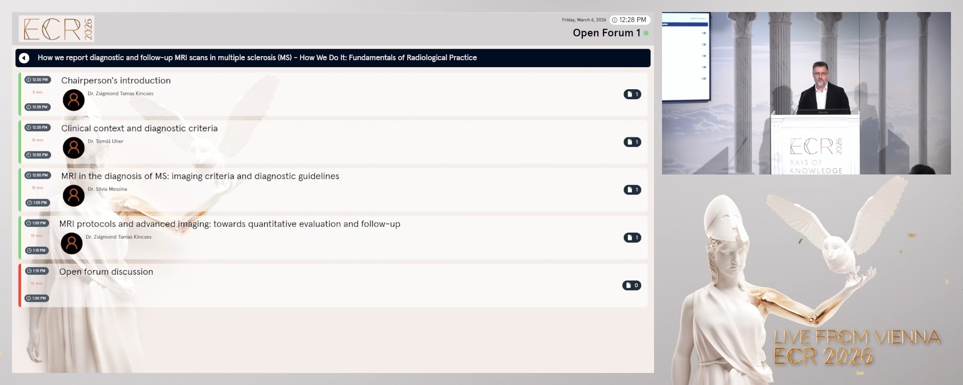
Chairperson's introduction
05:00Zsigmond Tamas Kincses, Szeged / HU
2

Clinical context and diagnostic criteria
15:00Tomáš Uher, Prague / CZ
3
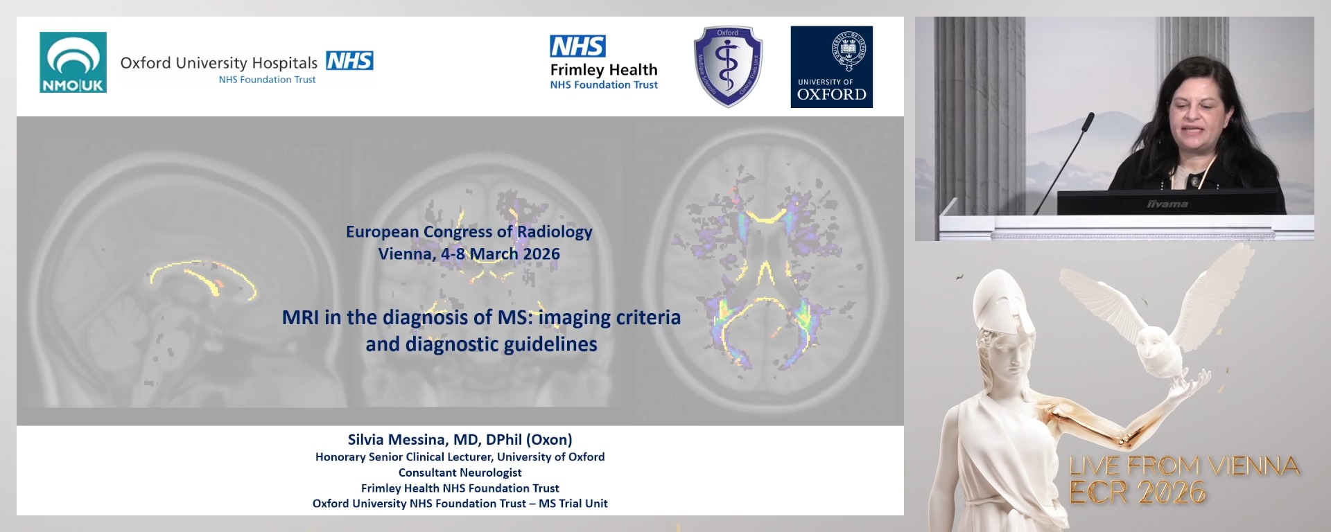
MRI in the diagnosis of MS: imaging criteria and diagnostic guidelines
15:00Silvia Messina, Oxford / UK
4
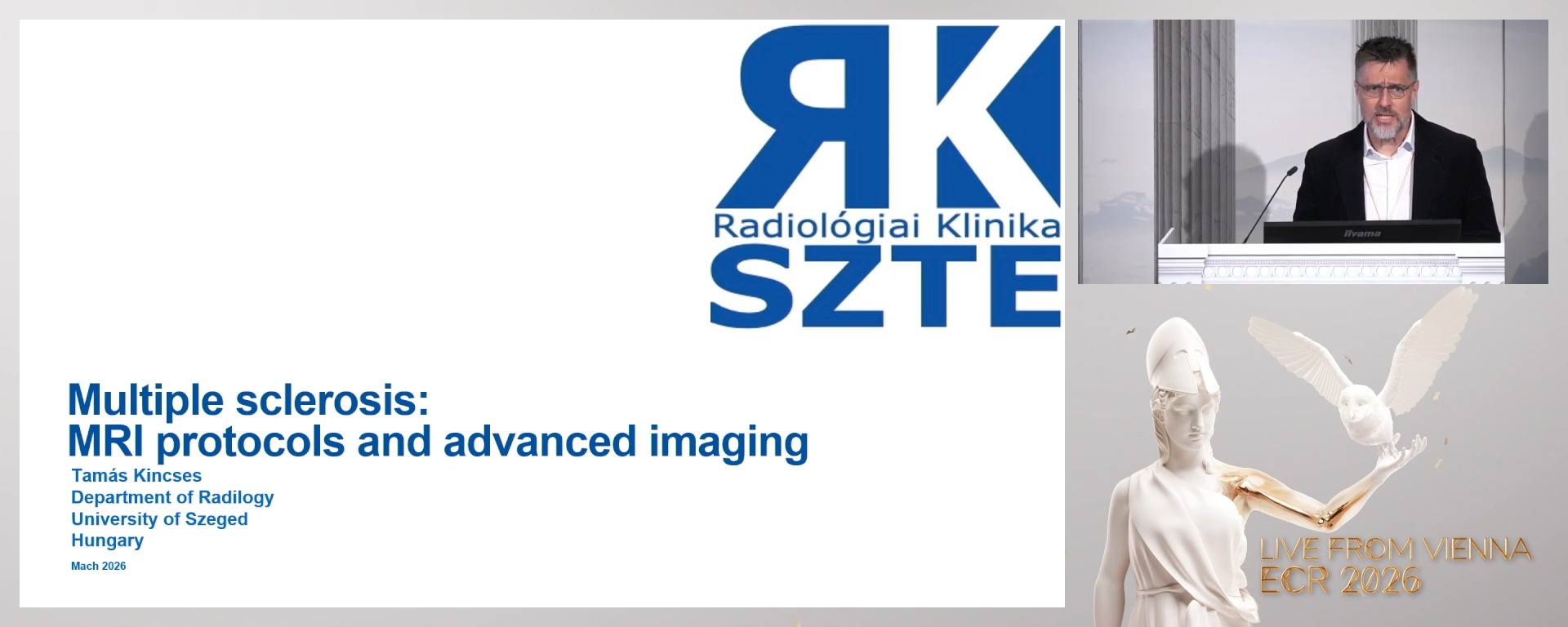
MRI protocols and advanced imaging: towards quantitative evaluation and follow-up
10:00Zsigmond Tamas Kincses, Szeged / HU
5
Open forum discussion
15:00Open forum discussion
5 min
Chairperson's introduction
Zsigmond Tamas Kincses, Szeged / Hungary
15 min
Clinical context and diagnostic criteria
Tomáš Uher, Prague / Czechia
- To explain the clinical presentation and differential diagnosis of multiple sclerosis and related disorders.
- To review the role of paraclinical tools (CSF, visual and somatosensory evoked potentials, etc.) and how they integrate with imaging in the diagnostic workflow.
- To learn how to apply the 2024 McDonald criteria with special attention to dissemination in space and time, RIS/CIS cases.
15 min
MRI in the diagnosis of MS: imaging criteria and diagnostic guidelines
Silvia Messina, Oxford / United Kingdom
- To explain the MRI features characteristic of MS lesions in the brain and spinal cord, including dissemination in space and time.
- To review international new diagnostic imaging guidelines and their application in clinical practice.
- To learn how to apply standardised reporting frameworks for MS, including lesion descriptors, anatomical localisation, terminology, and structured report templates.
10 min
MRI protocols and advanced imaging: towards quantitative evaluation and follow-up
Zsigmond Tamas Kincses, Szeged / Hungary
- To explain the recommended MRI acquisition protocols for MS diagnosis and follow-up, covering sequence selection, resolution, anatomical coverage, and harmonisation.
- To review advanced imaging techniques (e.g. volumetry, DTI, MTR, fMRI) and their potential role in monitoring disease progression and treatment response.
- To learn how to report diagnostic and follow-up MRI scans in MS using consistent language, longitudinal comparisons, and standardised criteria for new/enlarging lesions and atrophy.
15 min
Open forum discussion