In Focus: The Art of Artificial Intelligence in Clinical Practice
IF 18 - From snapshots to a story: Al in follow-up imaging after oncologic treatment
Lectures
1
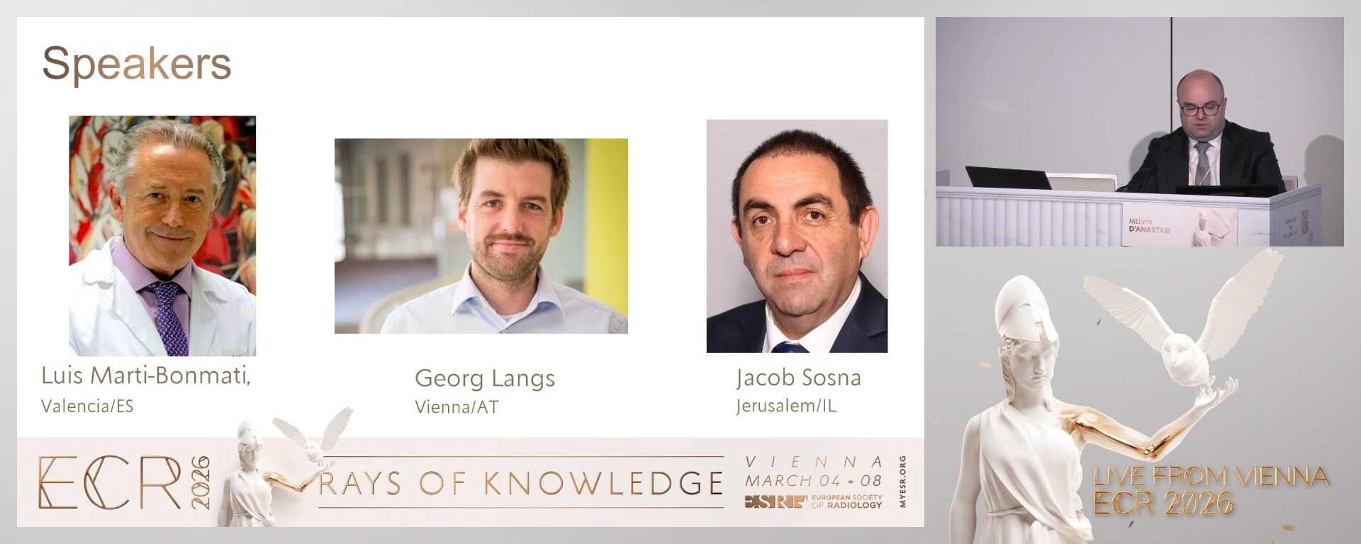
Chairperson's introduction
05:00Melvin D'Anastasi, Mosta / MT
2
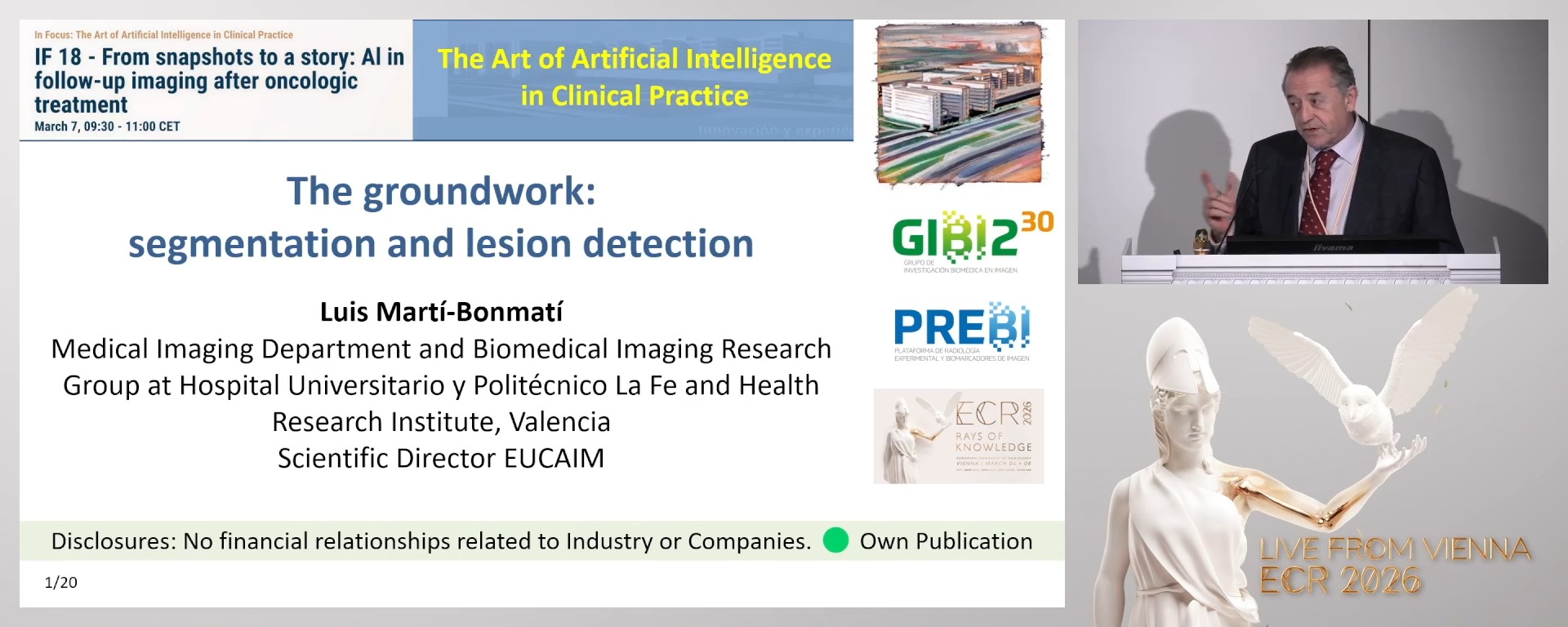
The groundwork: segmentation and lesion detection
20:00Luis Marti-Bonmati, València / ES
3
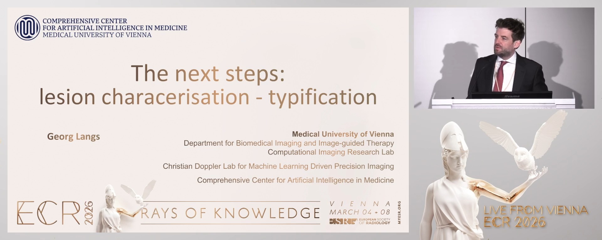
The next steps: lesion characterisation - typification
20:00Georg Langs, Vienna / AT
4
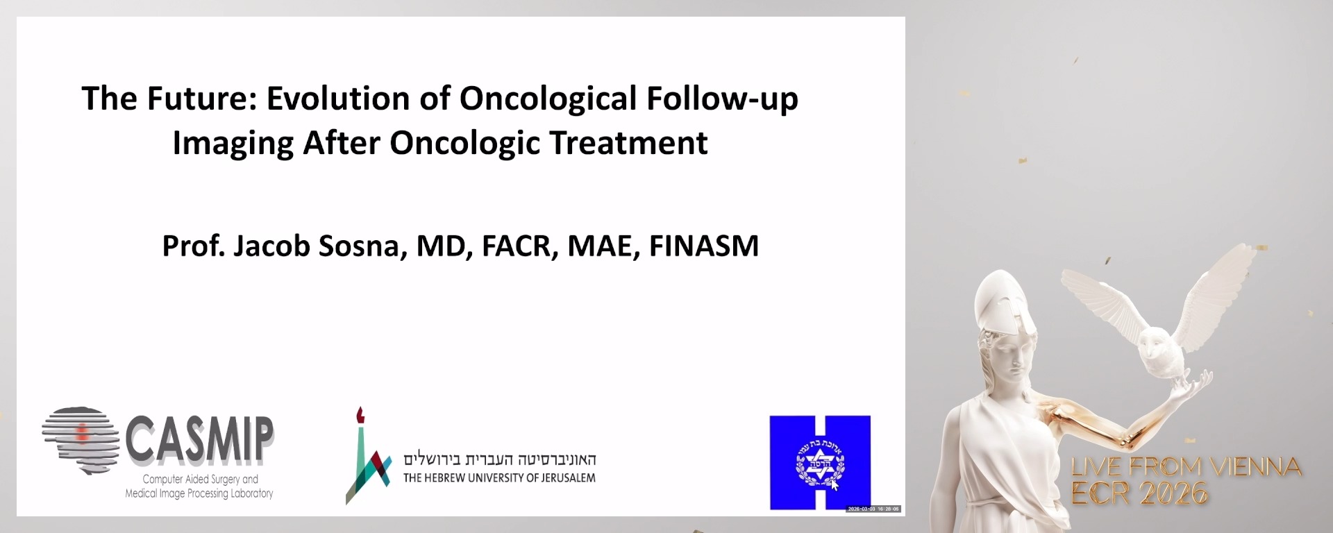
The future: evolution of oncologic follow-up imaging assessments
20:00Jacob Sosna, Jerusalem / IL
5
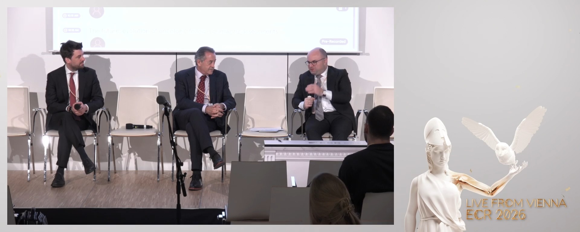
Panel discussion: When will AI automate manual work in oncologic imaging?
25:00Panel discussion: When will AI automate manual work in oncologic imaging?
Regular follow-up imaging is a cornerstone of oncologic care, enabling the monitoring of disease progression and treatment response. In radiology, this process involves the structured acquisition of comparable imaging studies over time - these individual exams are complete in their meaning based on their temporal relationship to one another. This session explores how Al already helps radiologists by enhancing and optimising oncologic follow-up imaging and how it can facilitate a more holistic assessment of oncological patients.
5 min
Chairperson's introduction
Melvin D'Anastasi, Mosta / Malta
20 min
The groundwork: segmentation and lesion detection
Luis Marti-Bonmati, Valencia / Spain
20 min
The next steps: lesion characterisation - typification
Georg Langs, Vienna / Austria
20 min
The future: evolution of oncologic follow-up imaging assessments
Jacob Sosna, Jerusalem / Israel
25 min
Panel discussion: When will AI automate manual work in oncologic imaging?