E³ - Young ECR Programme: Basic Session - Organised by the ESR
BS 18a - Intervertebral disc pathology: herniations, bulges and modic changes
Lectures
1
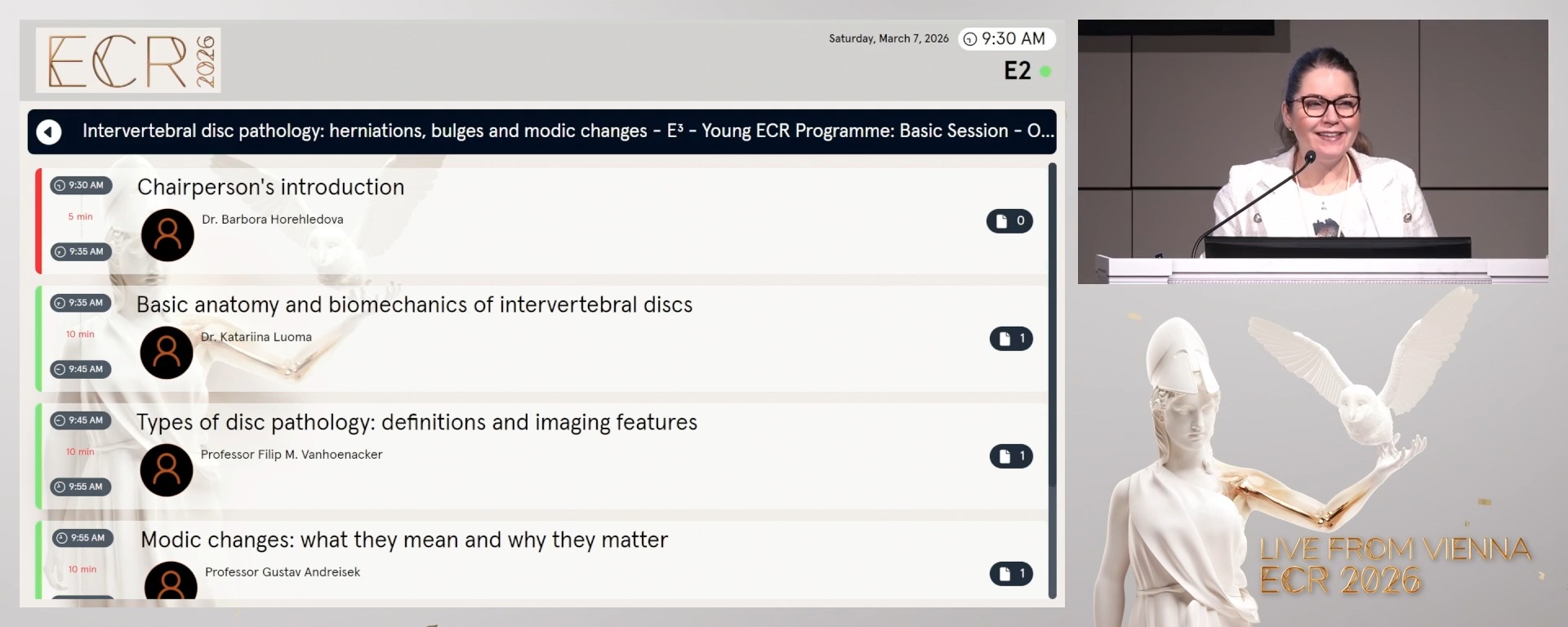
Chairperson's introduction
05:00Barbora Horehledova, Heerlen / NL
2
Basic anatomy and biomechanics of intervertebral discs
10:00Katariina Luoma, HHelsinki / FI
3
Types of disc pathology: definitions and imaging features
10:00Filip M. Vanhoenacker, Mechelen / BE
4
Modic changes: what they mean and why they matter
10:00Gustav Andreisek, Münsterlingen / CH
5
Case-based learning: real MRI examples - reporting tips for young radiologists
25:00Thomas Le Corroller, Marseille / FR
5 min
Chairperson's introduction
Barbora Horehledova, Heerlen / Netherlands
10 min
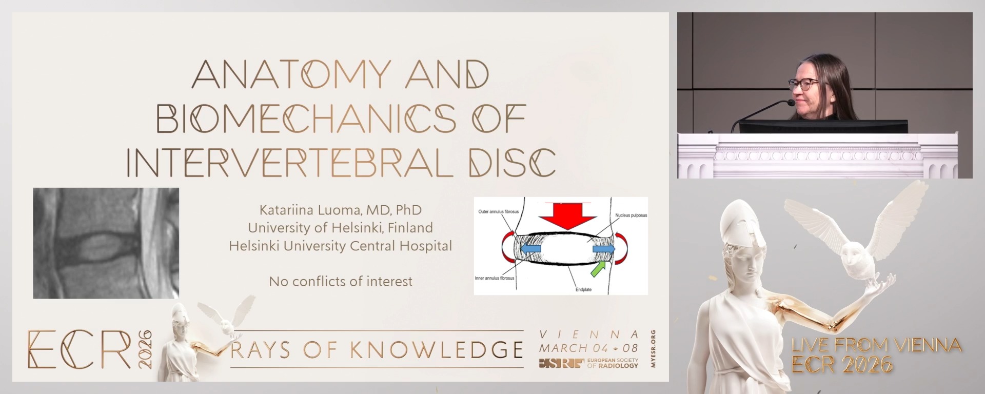
Basic anatomy and biomechanics of intervertebral discs
Katariina Luoma, HHelsinki / Finland
- To present the intervertebral disc structure: nucleus pulposus, annulus fibrosus, endplates.
- To explain the role in spinal stability and motion.
10 min
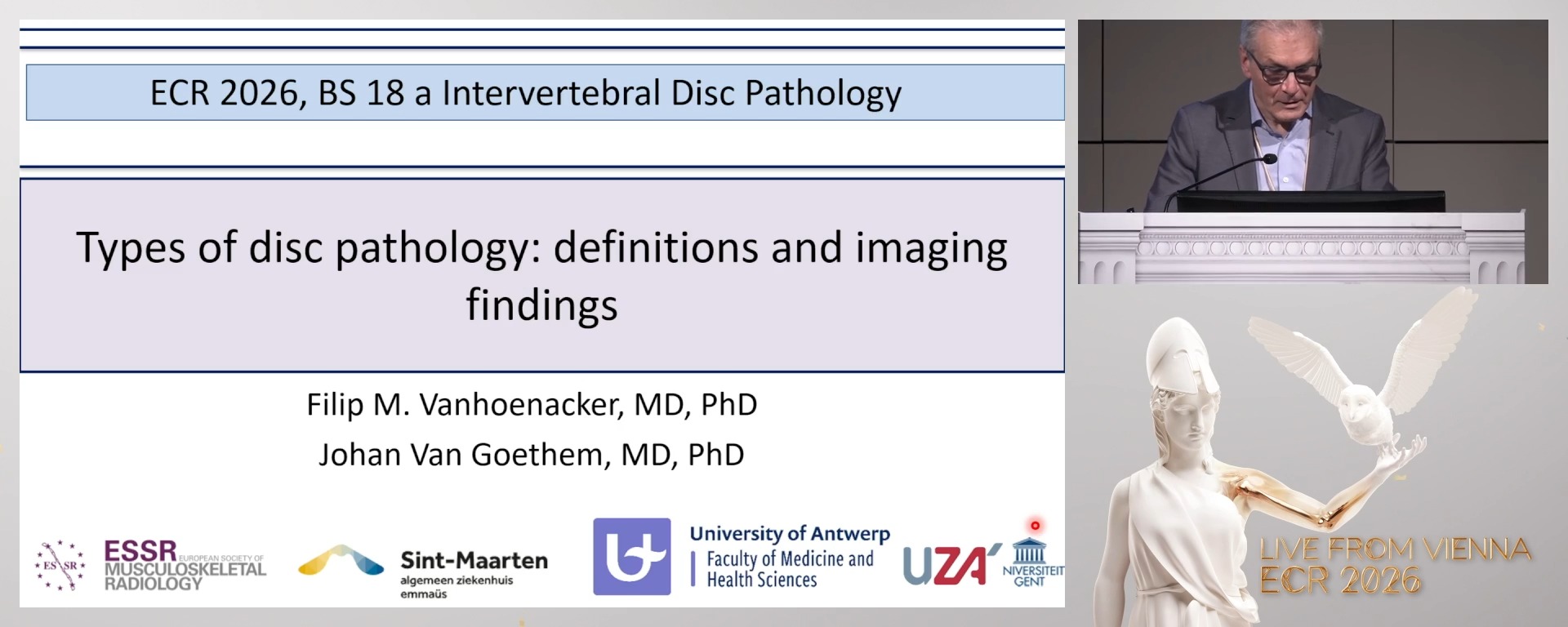
Types of disc pathology: definitions and imaging features
Filip M. Vanhoenacker, Mechelen / Belgium
- To differentiate between disc bulge vs herniation on MRI.
- To present T2-weighted MRI findings between disc protrusion vs extrusion vs sequestration.
- To correlate clinical relevance with symptoms.
10 min
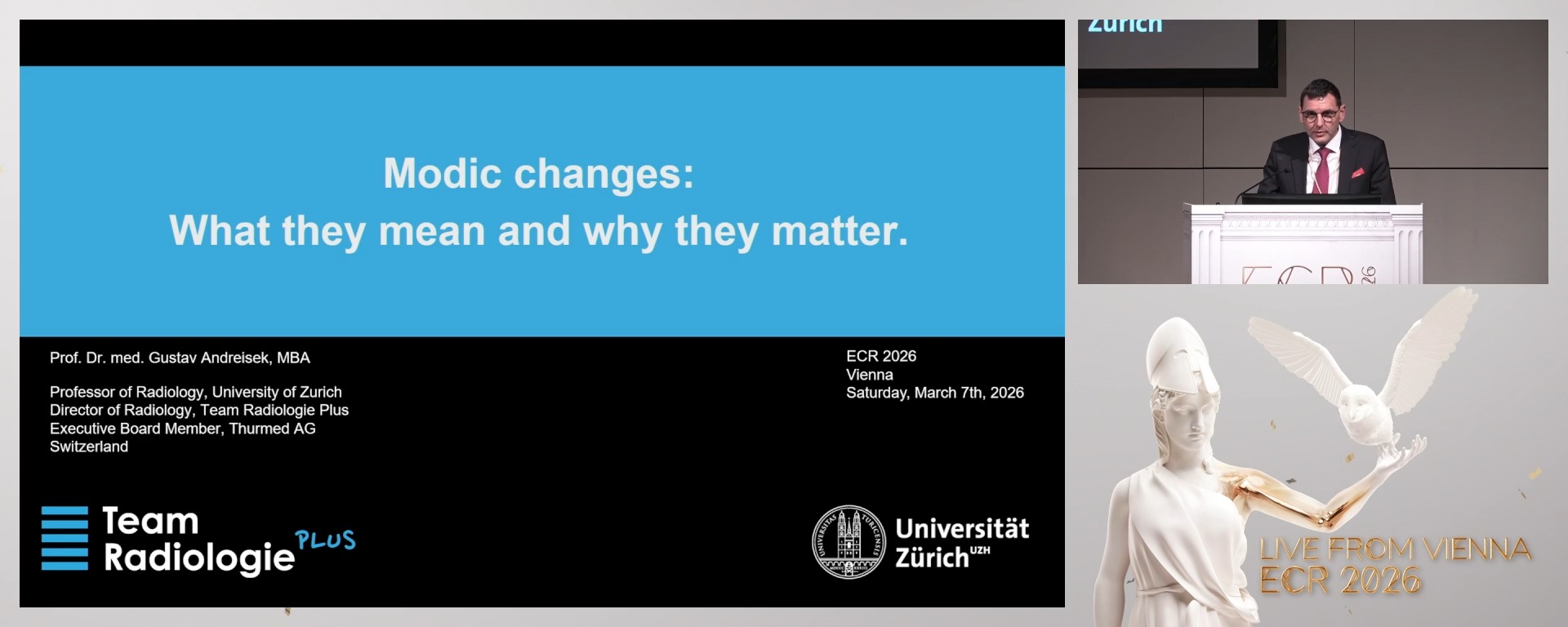
Modic changes: what they mean and why they matter
Gustav Andreisek, Münsterlingen / Switzerland
- To present type 1: edema/inflammation (T2 hyperintense, T1 hypointense).
- To present type 2: fatty degeneration (T1 hyperintense, T2 isointense).
- To present type 3: sclerosis (T1 and T2 hypointense).
- To correlate with degenerative disc dis
25 min
Case-based learning: real MRI examples - reporting tips for young radiologists
Thomas Le Corroller, Marseille / France
- To discuss interactively common findings.
- To recognise pitfalls and mimics such as infections and tumours.
- To present standardised terminology such as Fardon and Milette classification.
- To recommend when further imaging or intervention is needed.