Research Presentation Session: Radiographers
RPS 2214 - Radiographers' roles in angiography and interventional procedures
Lectures
1
Establishing Diagnostic Reference Levels (DRLs) for Neurointerventional Procedures in Malta
06:00Marvin Grech, Ghaxaq / MT
2
Saving lives and preventing harm in Jersey: Radiographer-Led Nasogastric Tube Position Check Pathway Pilot
06:00Gavin Cain, St. Helier / UK
3
Enhanced Arterial Imaging with Reduced Contrast Load: A Longitudinal Study of Weight-Based CTA Protocol Implementation
06:00Hortência De Jesus Ferreira, Campinas / BR
4
Predictive Modeling and Image Quality Metrics for Dose Reduction in CT-Guided Liver Ablation
06:00Tim Antheunis, Veenendaal / NL
5
Expanding radiographers’ knowledge and skills through Virtual Reality (VR): A bridging role in preoperative assessment. A pilot experience
06:00Andrea Masperi, Abbiategrasso / IT
6
Evaluation of Needle Artefact Reduction Techniques in CT-Guided Biopsy Imaging
06:00Clare Rainey, Cork / IE
7
Shaping Radiography Education: Radiographers Designing Modular 3D-Printed Angiographic Simulators
06:00Andrea Masperi, Abbiategrasso / IT
8
Radiation Dose Monitoring in Interventional Cardiology: Patient, Occupational, and Pediatric Perspectives from Latin America
06:00Marcus Vinicius Linhares Oliveira, Salvador / BR
9
Personalized Weight-Tiered Protocols in Angiotomography: The Synergy Between Canon Prime SP and Stellant SmartFlow Injector in Improving Diagnostic Quality
06:00José Thiago De Souza De Castro, Campinas / BR
6 min
Establishing Diagnostic Reference Levels (DRLs) for Neurointerventional Procedures in Malta
Marvin Grech, Ghaxaq / Malta
Author Block: M. Grech, F. Zarb, R. Grech, P. Bezzina; Msida/MT
Purpose: Interventional neuroradiology procedures are vital for managing neurological conditions but expose patients to ionising radiation. The aim of the study was to assess the level of radiation doses during diagnostic and therapeutic Neurointerventional procedures in Malta and to establish and compare local Dose Reference Levels (DRLs) with international standards.
Methods or Background: Retrospective data were collected from DoseWatch for the period January 2020 to May 2025, focusing on Air Kerma Area Product (PKA), Reference Air Kerma (RAK), and Fluoroscopy Time (FT) for the following procedures: Cerebral Angiography (CA), Stroke Thrombectomy (ST), Aneurysm Coiling (AC), and AVM/AVF embolization. DRLs were calculated as the 3rd quartile (75th percentile) of the dose distributions.
Results or Findings: In Malta, the 3rd quartile values for CA were considerably lower than those reported in the literature, with PKA at 58 Gycm², RAK at 330 mGy, and FT at 9 minutes. ST revealed radiation doses approximately half of those reported in the literature (PKA: 70 Gycm²; RAK: 372 mGy; FT: 23 minutes). For AC, the doses were comparable to international standards (PKA: 182 Gycm²; RAK: 3384 mGy; FT: 45 minutes). Conversely, radiation doses for AVM/AVF embolisation were considerably higher (PKA: 1463 Gycm²; RAK: 7900 mGy; FT: 168 minutes).
Conclusion: The findings emphasise the importance of understanding radiation exposure to enhance patient safety. Establishing local DRLs will provide benchmarks for practitioners in Malta, while enabling continuous monitoring and development of optimisation strategies to mitigate risks associated with ionising radiation.
Limitations: This was a single center study establishing local DRLs and comparing these to literature findings. The center where study took place is however the only center providing neurological services in Malta.
Funding for this study: No funding required
Has your study been approved by an ethics committee? Yes
Ethics committee - additional information: Ethical approval was sought and obtained from the University of Malta Research and Ethics Committee (UREC): (FHS-2024-00575)
6 min
Saving lives and preventing harm in Jersey: Radiographer-Led Nasogastric Tube Position Check Pathway Pilot
Gavin Cain, St. Helier / United Kingdom
Author Block: G. Cain1, N. Aslam1, D. J. M. Tolan2, G. Roe2, L. Horder3, A. Gill4; 1St. Helier, Jersey (Channel Islands)/UK, 2Leeds/UK, 3London/UK, 4Oxford/UK
Purpose: To evaluate the safety, timeliness, and accuracy of a radiographer-led nasogastric (NG) tube position check pathway pre- and post-implementation.
Methods or Background: A collaborative initiative involving professional bodies, including the Society and College of Radiographers and the Royal College of Radiologists, launched a national radiographer-led NG tube pathway. This empowers radiographers to assess NG tube placement on chest X-rays and take corrective action if tubes are misplaced. It’s a response to repeated safety alerts related to the inadvertent use of misplaced NG tubes—classified as ‘Never Events’ by the NHS. On 30th June 2025, Jersey General Hospital became the first pilot site in the UK and Channel Islands to implement the pathway.
A retrospective audit compared the first 12 weeks post-implementation (radiographers performing first-line placement checks) with a pre-implementation period (radiologist-only X-ray reporting). Metrics assessed included time from image acquisition to clinical evaluation, misplacement detection rate, corrective action on misplaced tubes, and concordance between radiographer evaluations and the definitive radiology report. Radiographers completed a national competency-based e-learning programme supported by governance and a Standard Operating Procedure.
Results or Findings: Median turnaround time for clinical evaluation was reduced from 242.2 hours (10.1 days) to 12 minutes post-implementation. Radiographers detected 100% of misplaced NG tubes (12/12) and took corrective action on all misplaced tubes identified in the department (6/6). Concordance with the definitive radiology report was 96.8% (60/62).
Conclusion: The radiographer-led pathway is safe and effective, significantly reducing delays and minimising the risk of Never Events. The Jersey pilot reinforces evidence that with appropriate training and governance, radiographers can enhance NG tube safety through assessment and action at the point of image acquisition.
Limitations: Small sample size; focused on radiographer accuracy rather than patient outcome.
Funding for this study: No
Has your study been approved by an ethics committee? Not applicable
Ethics committee - additional information:
6 min
Enhanced Arterial Imaging with Reduced Contrast Load: A Longitudinal Study of Weight-Based CTA Protocol Implementation
Hortência De Jesus Ferreira, Campinas / Brazil
Author Block: J. T. d. S. d. Castro, H. d. J. Ferreira, D. C. Novais da Silva, D. Yamada, S. San Juan Dertkigil, F. Reis; Campinas - Sao Paulo/BR
Purpose: Weight-based personalisation of contrast media in CTA can maintain or increase arterial enhancement while reducing contrast load; longitudinal real-world evidence remains scarce. The aim is to quantify the impact of weight-stratified CTA protocols on arterial enhancement and iodine use in a real-world setting.
Methods or Background: Single-centre retrospective longitudinal study within ANGIO-MONITOR. Consecutive angiographic CTAs were analysed across two periods: pre-implementation (fixed protocols; N=1,626) and post-implementation (from 01-Jun-2025; weight-based volumes/flows and PSI ceilings; N=558). Primary endpoint: arterial attenuation (HU) in the target vessel per exam. Secondary endpoints: proportion of “optimal” studies (≥350 HU), contrast volume (mL/exam; mL/kg), and repeat-acquisition rate. Statistical tests: Welch’s test for means, proportion comparisons with 95% CIs; exam-level analyses with temporal stratification.
Results or Findings: Arterial enhancement increased from 315.5 HU (95% CI 311.1–319.9) to 484.4 HU (469.8–499.1); p=7.1×10⁻⁷⁹. Mean contrast volume decreased from 79.5 mL (78.3–80.7) to 67.9 mL (65.8–69.9); p=1.1×10⁻²⁰. mL/kg fell from 1.09 (1.07–1.11) to 0.91 (0.88–0.94); p=5.8×10⁻²⁵. The proportion of optimal studies rose from 30.0% to 76.5% (absolute Δ +46.6 pp; RR 2.55; p<10⁻¹⁶). Repeat-acquisition rates were low and similar (1.91% vs 2.15%; p=0.72). Effects were observed across angiographic indications in routine operations.
Conclusion: Weight-based CTA protocols, implemented under technologist-led governance and injector/CT synchronisation, substantially increased arterial enhancement while reducing contrast exposure in real-world practice. Findings support institutional scale-up, underpin iodine stewardship and cost savings, and provide a foundation for indication-specific analyses and prospective predictive modelling.
Limitations: The study is a single-center retrospective analysis, which may limit the generalizability of the findings.
Funding for this study: Not Applicable
Has your study been approved by an ethics committee? Yes
Ethics committee - additional information: The work has been submitted to the university's Research Ethics Committee (CEP) and is currently under review for evaluation and final opinion.
6 min
Predictive Modeling and Image Quality Metrics for Dose Reduction in CT-Guided Liver Ablation
Tim Antheunis, Veenendaal / Netherlands
Author Block: T. Antheunis1, M. Hakkert2, B. Vermolen1; 1Ede/NL, 2Haarlem/NL
Purpose: CT-guided liver ablation is a minimally invasive procedure associated with substantial radiation exposure for both patients and staff. Current protocols are not optimized, resulting in unnecessary dose. This study aimed to achieve dose reduction through protocol optimization, validated by a predictive model correcting for clinical factors.
Methods or Background: A single-center interventional study was conducted at Ziekenhuis Gelderse Vallei. Group A (n=76) underwent ablation before protocol adjustment; Group B (n=7) after implementation. A linear regression model was developed from Group A to predict dose-length product (DLP) based on significant clinical factors (patient size, number of lesions). In parallel, a phantom study evaluated the impact of slice thickness, tube current, and reconstruction technique (hybrid iterative reconstruction vs. model based reconstruction) on task-based image quality using the detectability index (d’). The optimized protocol was applied to Group B and validated against model predictions.
Results or Findings: Patient size (r=0.76; p<0.001) and number of lesions (p=0.016) were the strongest dose determinants. The model explained 59% of DLP variance (RMSE=532 mGy·cm). Phantom analysis showed model based reconstructions maintained higher d’ at low dose compared to iDose (at 2 mGy: IMR=4.71 vs. iDose=1.66). The new protocol reduced CTDIvol for spiral scans to 2–4 mGy and differentiated between planning and control scans. Group B achieved a 40–70% DLP reduction versus predicted values, with clinically acceptable image quality.
Conclusion: Task-based image quality analysis combined with predictive modeling enables substantial radiation dose reduction without compromising diagnostic utility. The integration of IMR, reduced tube current, and adjusted slice thickness was key. This approach provides a robust framework for task-specific CT protocol optimization and is transferable to other interventional procedures.
Limitations: The limitations of the study are single centre study and
Funding for this study: No funding was received for this study.
Has your study been approved by an ethics committee? Not applicable
Ethics committee - additional information:
6 min
Expanding radiographers’ knowledge and skills through Virtual Reality (VR): A bridging role in preoperative assessment. A pilot experience
Andrea Masperi, Abbiategrasso / Italy
Author Block: A. Masperi, G. Di Stefano, C. Gallotta, M. Vertemati; Milan/IT
Purpose: To evaluate the value of 3D CT reconstructions visualised in VR for preoperative planning in paediatric and adult thoracic surgery, and to assess through a pilot study the role of radiographers as key professionals in the workflow, from image acquisition to VR scenario creation and clinical collaboration.
Methods or Background: We conducted a feasibility study including eight patients, five paediatric and three adults, selected in collaboration with the surgical team to represent different anatomical and pathological scenarios.
CT datasets were processed into patient-specific 3D models and integrated into immersive VR environments. The workflow included: 1) segmentation with 3D Slicer, 2) rendering optimisation in Blender, 3) VR development in Unity, and 4) deployment on Meta Quest 3. Models were validated with surgeons through a mixed assessment: a seven-item Likert survey (1–5) and open-ended feedback on anatomical accuracy and surgical planning.
Results or Findings: In paediatric cases, VR improved vascular and bronchial assessment, supporting thoracoscopic or conservative strategies. In adults, it enhanced anatomical understanding and team communication, with limited impact on surgical choices. All models scored highly (4–5/5) for accuracy and clinical value.
Conclusion: Radiographers demonstrated the ability to manage the full workflow from imaging to VR, enhancing data optimisation, 3D modelling, and teamwork to support surgical planning. By streamlining processes and reinforcing clinical decision-making, they establish themselves as key professionals in precision surgery.
Limitations: This study involved a small and heterogeneous cohort without a control group and relied on qualitative data. Model accuracy depended on CT quality, while the VR workflow required specific skills that were progressively acquired during the process. Nevertheless, it represents the first Italian experience of VR-based surgical planning led by radiographers.
Funding for this study: This study is based on the ongoing agreement for the provision of virtual 3D models between PRINTMED-3D (Integrated Platform for Three-Dimensional Medical Technologies), awarded in 2019 under the “Call Hub Research and Innovation” program by Regione Lombardia, supported within the MUSA (Multilayered Urban Sustainability Action) project funded in 2022 by the National Recovery and Resilience Plan (PNRR), and the Vittore Buzzi Children’s Hospital, part of ASST FBF-Sacco.
Has your study been approved by an ethics committee? Not applicable
Ethics committee - additional information:
6 min
Evaluation of Needle Artefact Reduction Techniques in CT-Guided Biopsy Imaging
Clare Rainey, Cork / Ireland
Author Block: S. Toomey, B. Normoyle, A. England, C. Rainey, N. Moore, L. McLaughlin, M. F. Mcentee, M. Maher; Cork/IE
Purpose: CT-guided lung biopsy plays an important role in diagnosing malignant lung lesions. However, the presence of biopsy needle containing metal components introduces unwanted streak artefact that can obscure needle tip visualisation. This study aims to evaluate the impact of various protocols and needle size on axial streak artefacts.
Methods or Background: Two needles 18G and 20G with and without stylets were imaged in a watermelon using protocols at 70,80,100,120 kV with multiple reconstruction techniques including ASIR, DLIR and MAR. Signal-to-noise ratio (SNR) and contrast-to-noise ratio (CNR) were calculated across multiple ROIs to objectively compare image quality. Line plot profiles were used to calculate the artefact width for each protocol.
Results or Findings: The Interventional Protocol produced the highest SNR values for 18G and 20G biopsy needle. In line plot profile analysis, less streak artefact from the biopsy needle was produced in the Interventional Protocol. In the 20G smaller gauge needle, produced less streak artefact with the stylet out compared to the 18G. The higher tube voltages particularly in 100 and 120 kV reduced streak artefact for both biopsy needles.
Conclusion: The Interventional Protocol demonstrated consistent metal artefact reduction generated by 18G and 20G biopsy needle. This reduction may lead to a better depiction of the needle tip inside the nodule of interest.
Limitations: Study acquisitions were based on imaging a watermelon, watermelons have been used in similar experiments but are not necessarily a valid substitute for humans,.
Funding for this study: None
Has your study been approved by an ethics committee? Yes
Ethics committee - additional information: Medical School Social Research Ethics Committee - University College Cork
6 min
Shaping Radiography Education: Radiographers Designing Modular 3D-Printed Angiographic Simulators
Andrea Masperi, Abbiategrasso / Italy
Author Block: A. Masperi, F. Cavaliere, S. Boniardi, M. Vertemati, F. Pezzotta; Milan/IT
Purpose: To pioneer a shift in radiographer education by moving beyond traditional lectures toward immersive, practice-based learning through simulation. Within the specialized field of interventional and angiographic radiology, radiography students conceived and built a modular 3D-printed angiographic simulator.
Methods or Background: A modular 3D-printed angiographic simulator was developed from angio-CT datasets extending from femoral to supra-aortic arteries. The workflow included: 1) image segmentation with 3D Slicer, 2) mesh refinement in Meshmixer, 3) modeling and support design with Fusion, 4) resin-based additive printing of vascular structures, and 5) post-processing and casting to assemble the simulator. Validation was conducted by radiographers and interventional radiologists using a 5-point Likert scale to assess anatomical fidelity and suitability for practice.
Results or Findings: Five radiographers and three interventional radiologists evaluated the 1:1 scale simulator, reporting high anatomical fidelity (median rating ≥4/5) and strong suitability for practicing catheter navigation and angiographic techniques, highlighting its potential as a safe and reproducible training tool.
Conclusion: This project established an innovative educational paradigm in interventional radiology through the creation of a modular 3D-printed angiographic simulator, designed by radiographers for radiographers. Beyond enhancing technical competence, critical reasoning, and professional autonomy, the simulator represents a scalable and reproducible model that can transform radiographer education internationally, fostering a new generation of professionals equipped for the evolving challenges of interventional practice.
Limitations: This study has some limitations. High production and assembly costs may hinder broader implementation, while radiographers’ skills in 3D printing and post-processing are still developing. Moreover, the simulator has not yet undergone structured testing with students, and its educational effectiveness requires further validation.
Funding for this study: None
Has your study been approved by an ethics committee? Not applicable
Ethics committee - additional information:
6 min
Radiation Dose Monitoring in Interventional Cardiology: Patient, Occupational, and Pediatric Perspectives from Latin America
Marcus Vinicius Linhares Oliveira, Salvador / Brazil
Author Block: M. V. L. Oliveira1, J. Ferreira1, I. Modesto1, E. Matos1, D. Andrade1, L. Santana1, C. Ubeda2, M. Navarro1; 1Salvador/BR, 2Arica/CL
Purpose: To review and synthesize recent Latin American research strategies on dose monitoring in fluoroscopy-guided and cardiology procedures, highlighting patient, occupational, and quality-assessment perspectives.
Methods or Background: A PubMed search was conducted using the terms Diagnostic reference level OR Radiation dose monitoring AND (fluoroscopy OR cardiology OR fluoroscopy guided interventional procedures) AND ("Latin America" OR Brazil OR Argentina OR Chile OR Colombia OR Peru OR Venezuela OR Ecuador OR Uruguay OR Paraguay OR Bolivia) for studies published between 2010and 2025. A thematic synthesis was applied to group studies into patient monitoring, risk assessment, pediatric DRLs, and occupational exposure.
Results or Findings: Twenty-two articles were selected. Brazilian institutions reported comprehensive patient dose registries, with one multicenter database covering over 10,000 cardiology procedures and identifying patients reaching substantial radiation dose levels (SRDLs), triggering dermatological follow-up. The MARP (Modelo de Avaliação de Risco Potencial) model compiled more than 200,000 records across 121 institutions, providing structured indicators for quality evaluation. Pediatric studies proposed initial DRLs using age- and weight-based criteria, addressing a critical gap in radiation protection for children. Occupational assessments revealed lens doses averaging 25–47 μSv/procedure, with estimated annual exposures of 6–11 mSv, and frequent extremity overexposures due to hand positioning.
Conclusion: Brazil’s initiatives illustrate the feasibility of structured dose monitoring and DRL implementation in interventional cardiology. Expanding these models through regional collaboration, standardized protocols, and occupational protection strategies is essential to strengthening radiation safety across Latin America.
Limitations: Coverage across Latin America remains limited, and gaps persist in multinational registries, prospective interventions, and long-term patient outcomes.
Funding for this study: Not applicable
Has your study been approved by an ethics committee? Not applicable
Ethics committee - additional information:
6 min
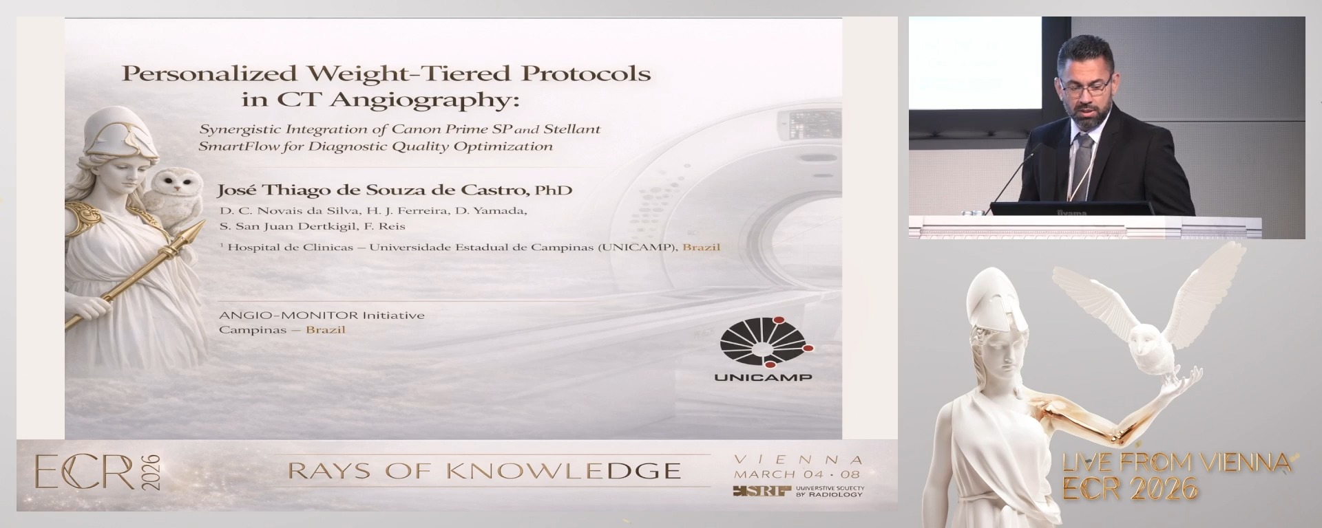
Personalized Weight-Tiered Protocols in Angiotomography: The Synergy Between Canon Prime SP and Stellant SmartFlow Injector in Improving Diagnostic Quality
José Thiago De Souza De Castro, Campinas / Brazil
Author Block: J. T. d. S. d. Castro, D. C. Novais da Silva, H. d. J. Ferreira, D. Yamada, S. San Juan Dertkigil, F. Reis; Campinas/BR
Purpose: Diagnostic quality in angiotomography relies on adequate vascular opacification (arterial Hounsfield Units, HU). Even with advanced technologies, standardized protocols ignoring patient weight often lead to suboptimal contrast. A study at HC-UNICAMP with 1,082 CTAs confirmed significant influence of weight, venous access type/caliber, contrast flow on arterial density. These findings informed personalized, weight-tiered protocols starting June 1, 2025.
Methods or Background: Retrospective analysis included 1,082 CTA exams performed with a Canon Prime SP 80-channel CT, Stellant SmartFlow injector (Bayer), and 300 mg/mL iodinated contrast. Variables collected included weight, age, venous access type/caliber, contrast flow, and arterial HU. Exams were quality-classified per ACR criteria: <250 HU (poor), 251–349 HU (average), and ≥350 HU (excellent). Following this analysis, new personalized weight-tiered protocols were implemented from June 2025, adjusting kVp, flow, contrast volume, and dose modulation for specific weight ranges (e.g., PE protocol for weights up to >100kg), with tailored parameters for each.
Results or Findings: Preliminary data shows significant improvement in contrast opacification: the percentage of exams >350 HU increased from 30% to 70%. An approximate 15% reduction in effective radiation dose was also observed. These are partial results from over 140 exams performed up to June 25, 2025, indicating that the personalized angiotomography protocols are extremely promising.
Conclusion: The implementation of personalized weight-tiered protocols in angiotomography at HC-UNICAMP marks a data-driven shift, optimizing diagnostic quality while reducing radiation dose and contrast volume. The synergy between the Canon Prime SP CT and Stellant SmartFlow injector, coupled with the radiology team's input, was crucial. This project serves as a model for other institutions, with prospective monitoring and full results to be presented at ESR2026.
Limitations: Results are being updated according to the new protocol's implementation, with ongoing data collection.
Funding for this study: Not applicable.
Has your study been approved by an ethics committee? Yes
Ethics committee - additional information: The submission to the Ethics Committee/IRB is currently under review