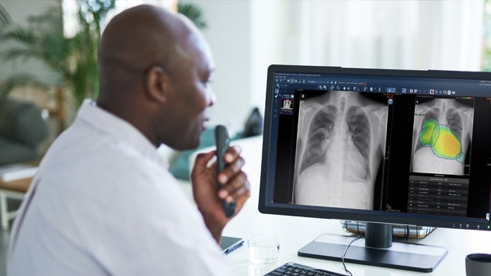
Chest X-ray AI: Taking on detection and diagnosis challenges in chest X-rays
Early detection of abnormal findings in the lungs can be critical to patients’ survival. Introducing the new RUBEE for AI package: Chest X-ray AI
They say extreme pressure creates diamonds, but that’s not what you are looking for to provide good patient care! The pressure on radiologists from the high volume of cases can, for example, create backlogs, make it difficult or impossible to prioritize cases, and even cause incidental findings to be missed.
While the Covid-19 pandemic has increased the demand for chest X-rays, the challenges of analyzing these critical images are not new. Looking for actionable nodules on the PACS or detecting tuberculosis in a chest image can be laborious and time-consuming, even for an expert.
Bringing nodules and lesions out of the shadows
Our newest RUBEE™ for AI curated package aims to take some of the pressure off analyzing chest X-rays.
Powered by Lunit INSIGHT CXR, and embedded in the Enterprise Imaging platform, Chest X-ray AI Analysis is a workflow-centric, evidence-based AI package that lightens the radiologist’s workload and reduces some of the challenges of detection and interpretation.
INSIGHT CXR can accurately detect 10 common findings in a chest X-ray and generates the analysis result which indicates the presence and the location of chest abnormalities.
Using abnormality scores, Chest X-ray AI Analysis can detect areas with suspicious findings and use deep learning to automatically analyze the images. It provides visualization and quantitative estimation of the presence of each abnormality, with a report that summarizes the overall analysis result, narrowed down to each finding.
Fast triage
‘Normal’ cases are triaged without any human intervention, freeing radiologists to focus on exams that are more likely to have an issue. Based on the abnormality score, the radiologist can then further prioritize which exams to read first.
And for triaging possible Covid-19 cases, Chest X-ray AI Analysis can help detect the presence of pneumonia quickly and accurately, so the patient can be isolated and begin treatment right away.
”17% Improved Nodule detection”
“19 % Faster Reads”
Improved diagnostic accuracy
The advanced visualization improves reading performance and supports any reader from a GP to a thoracic specialist to improve their diagnostic accuracy. It’s also easier to find those subtle incidental findings, such as small nodules that may be hidden by the hilar shadow, ribs, heart or diaphragm.
Finally, by improving diagnostic accuracy and speeding up reading time, Chest X-ray AI Analysis supports a smoother, faster emergency department workflow, with more informed decision-making that enables better patient care.

RUBEE™ for AI embeds AI seamlessly in Enterprise Imaging Workflows.
The Chest X-Ray AI Specialty Package is one of the AI Specialty packages that are available for RUBEE™ for AI.
The carefully selected, specialty-focused algorithms are seamlessly integrated in your Enterprise Imaging platform. Powered by RUBEE™ for AI in medical imaging, they can fully address your clinical, academic and research needs.
To learn how to turn AI into true Augmented Intelligence, visit the Agfa HealthCare website.
Cut through the AI hype to achieve real Augmented Intelligence results – a 30 min Virtual Lecture.
In recent months, Dr. Anjum M. Ahmed (MBBS, MBA, MIS), Agfa HealthCare’s Chief Medical Officer, has shared his insights and expertise on true Augmented Intelligence with leading healthcare providers across the globe, and is supporting them to cut through the AI hype.
With the right approach, and by embedding AI into clinical programs, AI becomes true Augmented Intelligence resulting in higher productivity and improved clinical confidence.
Hear from Anjum directly in this 30-minute Virtual Lecture, accessible at your convenience.
Access here

Dr. Anjum Ahmed, Chief Medical Officer Agfa HealthCare