Clinical Trials in Radiology
CTiR 18 - Clinical Trials in Radiology: cardiac, gastrointestinal and interventional studies
Lectures
1
Chairpersons' introduction
06:00Hatem Alkadhi, Zurich / CH, Ferdia Aidan Gallagher, Cambridge / UK
2
Subgroup analysis of Cardiac MRI in a phase 3 trial investigating safety and efficacy of gadoquatrane for low-dose contrast-enhanced MRI of the body
08:00Jeanette Schulz-Menger, Berlin / DE
3
Discussant
04:00Giles Hannibal Roditi, Glasgow / UK
4
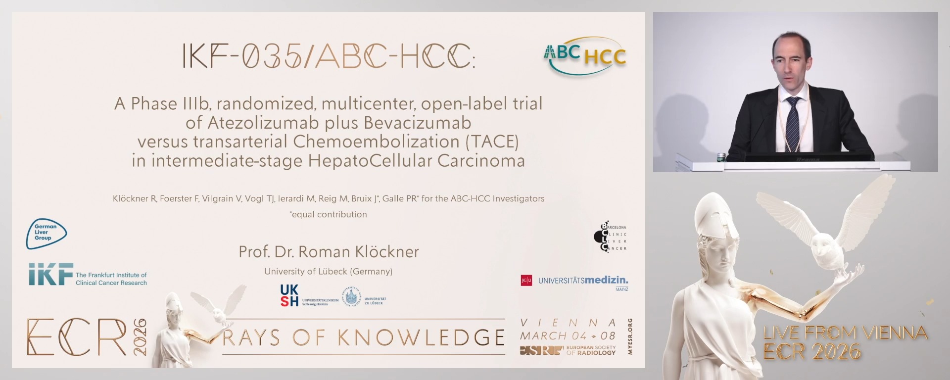
IKF-035/ABC-HCC: A Phase IIIb, randomized, multicenter, open-label trial of Atezolizumab plus Bevacizumab versus transarterial Chemoembolization (TACE) in intermediate-stage HepatoCellular Carcinoma
08:00Roman Klöckner, Lübeck / DE
5

Discussant
04:00Irene Bargellini, Candiolo / IT
6

Machine learning based assessment of the left atrium and left atrial appendage to predict atrial fibrillation in the SCOT-HEART trial
08:00Michelle Claire Williams, Edinburgh / UK
7
Discussant
04:00Karl-Friedrich Kreitner, Mainz / DE
8
Early Coronary CT Angiography for Non-Culprit Plaque Characterization After Primary PCI: Preliminary Results from the CT-STEMI Study
08:00Ludovica Blasi, Turin / IT
9
Discussant
04:00Milán Vecsey-Nagy, Budapest / HU
10
Prospective Multicentre Study investigating the management of patients with Malignant Ureteric Obstruction (OPTIMISE MUO) interim analysis of a multi-centre prospective observational study
08:00Oliver Llewellyn, Edinburgh / UK
11
Discussant
04:00Malgorzata Szczerbo-Trojanowska, Lublin / PL
12
Real-world implementation of objective motility MRI scoring in radiologist assessment of ileal Crohn’s disease on MRE: The CONTEXT Trial
08:00Iyad Naim, London / UK
13
Discussant
04:00Martina Scharitzer, Vienna / AT
6 min
Chairpersons' introduction
Ferdia Aidan Gallagher, Cambridge / United Kingdom
Hatem Alkadhi, Zürich / Switzerland
Hatem Alkadhi, Zürich / Switzerland
8 min
Subgroup analysis of Cardiac MRI in a phase 3 trial investigating safety and efficacy of gadoquatrane for low-dose contrast-enhanced MRI of the body
Jeanette Schulz-Menger, Berlin / Germany
4 min
Discussant
Giles Hannibal Roditi, Glasgow / United Kingdom
8 min
IKF-035/ABC-HCC: A Phase IIIb, randomized, multicenter, open-label trial of Atezolizumab plus Bevacizumab versus transarterial Chemoembolization (TACE) in intermediate-stage HepatoCellular Carcinoma
Roman Klöckner, Lübeck / Germany
Author Block: R. Klöckner1, F. Foerster2, V. Vilgrain3, T. Vogl4, A. M. Ierardi5, M. Reig6, J. Bruix6, P. R. Galle2, *. For the ABC-HCC Investigators7; 1Lübeck/DE, 2Mainz/DE, 3Clichy/FR, 4Frankfurt/DE, 5Milan/IT, 6Barcelona/ES, 7Frankfurt am Main/DE
Purpose: Transarterial chemoembolization (TACE) is standard of care (soc) for intermediate stage (BCLC B) HCC. IMbrave150 trial demonstrated that combining the anti-PD-L1 antibody atezolizumab and anti-VEGF antibody bevacizumab (atezo/bev) significantly improves overall survival compared to sorafenib as first-line treatment of advanced/intermediate stage HCC failing/unsuited for TACE. It remains unknown if atezo/bev is more efficacious than TACE in patients who would be treated with TACE per soc.
Methods or Background: Several trials assess the benefit of combining systemic therapy with TACE. In contrast, ABC-HCC, an international phase 3b, randomized, multicenter, open-label, investigator-initiated trial directly compares atezo/bev vs. TACE in intermediate stage HCC or HCC indicated for TACE according to treating physicians. In total, 320 patients with confirmed HCC (not amenable to curative surgery/ablation transplantation, no extrahepatic spread, no macrovascular invasion except Vp1/2, ECOG ≤1, Child-Pugh A/B7) are randomized (1:1) to receive atezo/bev (Arm A) or TACE (Arm B) for max 24 months. Imaging (CT/MRI, Q8W) determines the primary endpoint time to failure of treatment strategy (TTFS). Here, we report on the first of two interim analyses (IA) at 33% information time (85 events) assessing efficacy and futility.
Results or Findings: At data cut-off (13-Jun-2025), 194 patients were randomized at 54 centers in seven countries. Of these, 168 patients were included in the IA (A:87; B:81) and 100 events were observed for TTFS (A:44; B:56). Median TTFS was 14.6 months (A) vs. 9.5 months (B) with a HR of 0.55 (95% CI [0.36-0.83]).
Conclusion: The results provide first signals suggesting a superiority of atezo/bev compared to TACE in intermediate stage HCC. Therefore, the trial progresses to the second IA at 66% information time (169 events).
Limitations: The presented interim analysis was performed after the occurrence of only 33% of planned events.
Funding for this study: Funding for this study was provided by the legal sponsor IKF – The Frankfurt Institute of Clinical Cancer Research.
Additionally, the study is financially supported by F. Hoffmann-La Roche Ltd.
Has your study been approved by an ethics committee? Yes
Ethics committee - additional information: The study is approved in CTIS (EU CT No.: 2024-512953-26-00).
Written informed consent was obtained from all participants.
This abstract reports on original research.
Additional trial identifiers:
IKF No.: IKF-035
ClinicalTrials.gov: NCT04803994
EudraCT No.: 2020-004210-35
Roche No.: MO42581
AIO No.: AIO-HEP-0321/ass
4 min
Discussant
Irene Bargellini, Candiolo / Italy
8 min
Machine learning based assessment of the left atrium and left atrial appendage to predict atrial fibrillation in the SCOT-HEART trial
Michelle Claire Williams, Edinburgh / United Kingdom
4 min
Discussant
Karl-Friedrich Kreitner, Mainz / Germany
8 min
Early Coronary CT Angiography for Non-Culprit Plaque Characterization After Primary PCI: Preliminary Results from the CT-STEMI Study
Ludovica Blasi, Torino / Italy
Author Block: L. Blasi1, E. Puglisi1, T. D'Angelo2, M. Olivieri3, M. Moretti1, D. Verna1, R. Aroasio1, R. Faletti4, M. Gatti1; 1Turin/IT, 2Messina/IT, 3Chieti/IT, 4Candiolo/IT
Purpose: To assess burden and morphological characteristics of non-culprit plaques in STEMI patients undergoing early coronary CT angiography (CCTA) following PCI.
Methods or Background: 67 consecutive patients (mean age 62.5 ± 9.9 years; 52 men) were enrolled in the multicenter CT-STEMI study (NCT05941585). Coronary CT angiography was performed at a median of 9 days after the infarction [IQR 4]. All 16 coronary segments were evaluated per patient for the presence of stents, plaques, stenosis severity (scale 0–5), vulnerable plaque features (positive remodeling, low attenuation, spotty calcification, napkin-ring sign), Segment Involvement Score (SIS) and Segment Stenosis Score (SSS).
Results or Findings: Among 1072 segments analyzed, plaques were present in 33.2%, with 81.5% showing <50% stenosis, 12.9% moderate (50–69%), and 23 residual severe lesions (≥70% or occluded) in 15 patients, including 3 in proximal locations. A total of 148 segments contained stents. Overall, 173 plaques showed at least one high-risk feature: 105 with positive remodeling, 170 with low attenuation, 34 with spotty calcification, and 10 with napkin-ring sign. A single high-risk plaque (defined as having ≥2 features) was found in 17 patients (25.4%), while 30 patients (44.8%) had two or more. In proximal segments alone, 46 high-risk plaques were observed. The mean SIS was 5.3±2.6 and the mean SSS was 10.0±5.7.
Conclusion: Early CCTA in STEMI patients reveals a significant residual atherosclerotic burden, predominantly composed of plaques with mild to moderate stenosis. However, a notable number of severe and morphologically high-risk lesions, especially in proximal segments, were also detected. These findings, once correlated with clinical outcomes, may support more targeted therapeutic strategies.
Limitations: Limitations include small sample size, preliminary nature of the data, variable CT timing post-MI and segment-based analysis.
Funding for this study: This research was supported by a grant from the Italian Ministry of Health under the “Ricerca Finalizzata 2021 – Giovani Ricercatori” program, project number GR-2021-12372092. The funding was allocated to the project titled “Cardiac Computed Tomography for Comprehensive Risk Stratification of Arrhythmic, Atherothrombotic, and Heart Failure Events Following Reperfused ST-Segment Elevation Myocardial Infarction”. The funder had no role in the design, data collection, analysis, or interpretation of this study
Has your study been approved by an ethics committee? Yes
Ethics committee - additional information: No additional information
4 min
Discussant
Milán Vecsey-Nagy, Budapest / Hungary
8 min
Prospective Multicentre Study investigating the management of patients with Malignant Ureteric Obstruction (OPTIMISE MUO) interim analysis of a multi-centre prospective observational study
Oliver Llewellyn, Edinburgh / United Kingdom
Author Block: O. Llewellyn1, J. Blackmur1, J. Aning2, M. Bagkeris3, T. Barrett4, N. Shaida4, A. Laird1, U. Collaborative1, B. Oncology1; 1Edinburgh/UK, 2Bristol/UK, 3London/UK, 4Cambridge/UK
Purpose: Unlike other oncology emergencies such as metastatic spinal cord compression, no standardised care pathway exists for malignant ureteric obstruction (MUO) and there is geographical variation in management approach. This study aims to understand the rationale for management choices made by clinicians in patients with MUO and lay the groundwork for further work.
Methods or Background: National UK-wide multicentre observational prospective research study using national Interventional Radiology and Urology stakeholder networks (UNITE & BAUS). This is an HRA approved, RCR funded research study.
MUO patients will be identified by research teams from referrals (in and out of hours) and multidisciplinary team meetings. Pseudonymised data will be recorded using the REDCap platform. Data about reason for referral will be captured and patients will be followed up for 1 year at 3 month timepoints to capture survival, further treatment, and hospital re-admissions and re-interventions.
Results or Findings: We will present an interim analysis of this work which follows on from extensive previous multi-centre work (INSITE MUO) demonstrating an overall survival benefit for intervening (nephrostomy or ureteric stent) in advanced cancer involving the abdomen and causing malignant ureteric obstruction (p = 0.049). We suspect there are confounding factors influencing this finding and this study will allow us to present data addressing this.
Conclusion: Intervention (nephrostomy or ureteric stent) in MUO in advanced cancer appears to have a protective effect on overall survival. We will present interim findings of the OPTIMISE MUO study addressing reasons for this.
Limitations: Observational nature of methodology.
Funding for this study: Royal College of Radiologists, Kodak fellowship
Has your study been approved by an ethics committee? Not applicable
Ethics committee - additional information:
4 min
Discussant
Malgorzata Szczerbo-Trojanowska, Lublin / Poland
8 min
Real-world implementation of objective motility MRI scoring in radiologist assessment of ileal Crohn’s disease on MRE: The CONTEXT Trial
Iyad Naim, London / United Kingdom
4 min
Discussant
Martina Scharitzer, Vienna / Austria