Clinical Trials in Radiology
CTiR 20 - Late-breaking clinical trials in radiology
Lectures
1
Chairpersons' introduction
10:00Ferdia Aidan Gallagher, Cambridge / UK, James Alan Brink, Boston / US
2
Real-World Impact of AI Chest Radiograph Triage on Lung Cancer Pathways and Outcomes Across a UK NHS Trust
11:00Geraldine Dean, London / UK
3
Discussant
05:00Georg Langs, Vienna / AT
4
Quantitative cardiac CT at Photon Counting to unveil residual risk of rapid coronary atherosclerosis progression in stable patients undergoing optimal medical treatment
11:00Mariaelena Occhipinti, Pisa / IT
5
Discussant
05:00Ann-Christine Stahl, Berlin / DE
6
Association of Liver Fat Content with Major Adverse Cardiovascular Events in DISCHARGE and SCOT-HEART using the GUIDE-IT Data Sharing Platform
11:00Jakob Knape, Berlin / DE
7
Discussant
05:00Jonathan R. Weir-Mccall, London / UK
8
Diagnostic performance of an MRI-based AI device for Alzheimer’s disease: a multicentre retrospective study of synthetic CSF biomarkers
11:00Arnaud Attye, Grenoble / FR
9

Discussant
05:00Tomasz Matys, Cambridge / UK
10
Randomized trial of MRI-guided transurethral ultrasound ablation (TULSA) vs. radical prostatectomy for intermediate-risk prostate cancer: perioperative outcomes
11:00Katarzyna J. (Kasia) Macura, Baltimore / US
11

Discussant
05:00Sungmin Woo, New York / US
10 min
Chairpersons' introduction
Ferdia Aidan Gallagher, Cambridge / United Kingdom
James Alan Brink, Boston / United States
James Alan Brink, Boston / United States
11 min
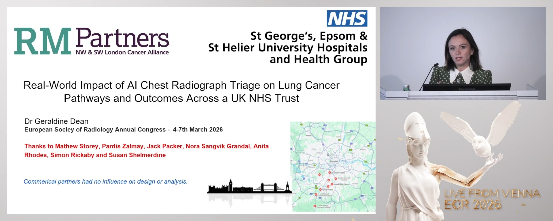
Real-World Impact of AI Chest Radiograph Triage on Lung Cancer Pathways and Outcomes Across a UK NHS Trust
Geraldine Dean, London / United Kingdom
Author Block: M. Storey, E. Antrum, J. Packer, P. Zalmay, G. Dean, S. C. Shelmerdine; London/UK
Purpose: To assess the real-world clinical impact and safety of artificial intelligence (AI) chest radiograph (CXR) triage following deployment across a large National Health Service (NHS) trust, addressing uncertainties regarding pathway acceleration and cancer stage shift.
Methods or Background: Three linked evaluations across five hospitals used a commercial AI system (Annalise CXR Enterprise v2.3): (1) a before–after analysis of 56,257 CXRs (2022–2024) assessing time to CT for suspected lung cancer after AI triage to a same-day CT pathway; (2) a prospective silent trial with failure analysis of normal CXR deprioritisation in 63,083 adults (2023); and (3) a longitudinal comparison of lung cancer investigations and stage before (2022) and after AI deployment (2023) with 12-month follow-up.
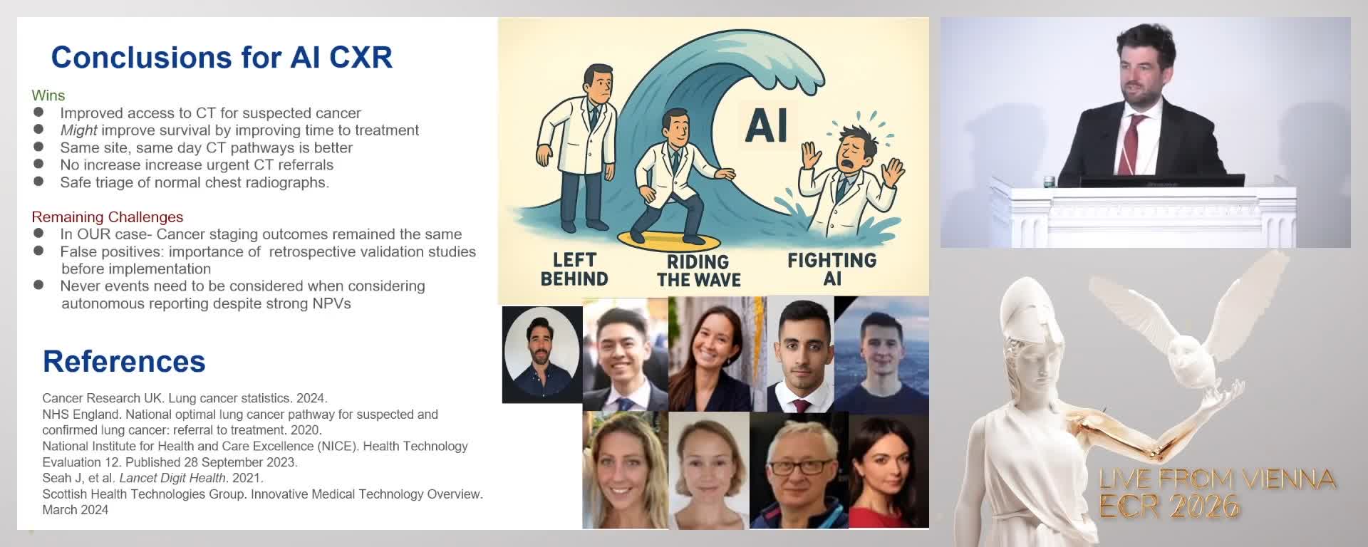
Results or Findings: Mean time from CXR to CT report for suspected lung cancer fell from 6.0 to 3.6 days (p<0.001), with greatest gains at sites with co-located CXR and CT (p<0.001), without increasing CT utilisation. The AI demonstrated 82.5% sensitivity and 90.5% specificity for cancer features. For normal radiographs, AI enabled potential deprioritisation of 18.5% of exams, with a clinically significant miss rate of 0.05%. Despite faster pathways, early-stage lung cancer diagnoses (13 pre-AI vs 11 post-AI) and investigation volumes (177 vs 160) remained unchanged.
Conclusion: AI CXR triage can be safely deployed to accelerate lung cancer diagnostic pathways and reduce reporting burden without increasing imaging volume but does not affect lung cancer stage distribution. These findings provide critical real-world evidence informing national guidance and the clinical debate on the downstream impact of AI in cancer care.
Limitations: Observational design with limited power to detect stage shift; overall survival not assessed. Results reflect a specific AI system and thresholds, and may not generalise. Normal CXR detection used a proxy reference standard.
Funding for this study: None
Has your study been approved by an ethics committee? Not applicable
Ethics committee - additional information:
Purpose: To assess the real-world clinical impact and safety of artificial intelligence (AI) chest radiograph (CXR) triage following deployment across a large National Health Service (NHS) trust, addressing uncertainties regarding pathway acceleration and cancer stage shift.
Methods or Background: Three linked evaluations across five hospitals used a commercial AI system (Annalise CXR Enterprise v2.3): (1) a before–after analysis of 56,257 CXRs (2022–2024) assessing time to CT for suspected lung cancer after AI triage to a same-day CT pathway; (2) a prospective silent trial with failure analysis of normal CXR deprioritisation in 63,083 adults (2023); and (3) a longitudinal comparison of lung cancer investigations and stage before (2022) and after AI deployment (2023) with 12-month follow-up.
Results or Findings: Mean time from CXR to CT report for suspected lung cancer fell from 6.0 to 3.6 days (p<0.001), with greatest gains at sites with co-located CXR and CT (p<0.001), without increasing CT utilisation. The AI demonstrated 82.5% sensitivity and 90.5% specificity for cancer features. For normal radiographs, AI enabled potential deprioritisation of 18.5% of exams, with a clinically significant miss rate of 0.05%. Despite faster pathways, early-stage lung cancer diagnoses (13 pre-AI vs 11 post-AI) and investigation volumes (177 vs 160) remained unchanged.
Conclusion: AI CXR triage can be safely deployed to accelerate lung cancer diagnostic pathways and reduce reporting burden without increasing imaging volume but does not affect lung cancer stage distribution. These findings provide critical real-world evidence informing national guidance and the clinical debate on the downstream impact of AI in cancer care.
Limitations: Observational design with limited power to detect stage shift; overall survival not assessed. Results reflect a specific AI system and thresholds, and may not generalise. Normal CXR detection used a proxy reference standard.
Funding for this study: None
Has your study been approved by an ethics committee? Not applicable
Ethics committee - additional information:
5 min
Discussant
Georg Langs, Vienna / Austria
11 min
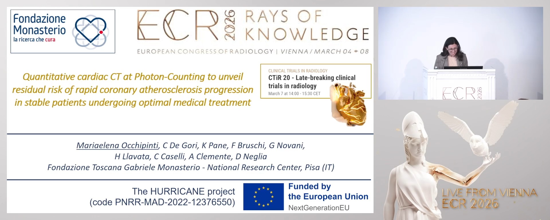
Quantitative cardiac CT at Photon Counting to unveil residual risk of rapid coronary atherosclerosis progression in stable patients undergoing optimal medical treatment
Mariaelena Occhipinti, Pisa / Italy
Author Block: M. Occhipinti1, C. De Gori1, K. Pane2, F. Brusch1, G. Novani1, H. Hlavata1, C. Caselli1, A. Clemente1, D. Neglia1; 1Pisa/IT, 2Naples/IT
Purpose: The prospective HURRICANE study (Health improvement by Understanding Residual Risk In CAd and NEw targets for treatment) aim to assess whether advanced quantitative cardiac computed tomography angiography (CCTA) may help identifying subjects at risk of rapid CAD progression despite optimal medical treatment (OMT) among patients with stable CAD.
Methods or Background: Among 221 patients with suspected CAD enrolled in the prospective HURRICANE study between May 2023 and November 2024, 93 patients (68±8yrs, 63%M) with complete 1 yr follow-up were selected. All patients had high quality CCTA at PCCT at baseline and follow-up (interscan period 344±44 days) and full clinical/biochemical characterization. CCTA exams were analysed according to CAD-RADS2 classification and by using a commercially available software program for semi-automated quantification of total plaque volume (PV), as well as non-calcified (NC, HU ≤350) and calcified (CA, HU>350) plaque components.
Results or Findings: The majority of patients (65%) were already treated with statins at baseline and statin use further increased at follow-up (78%). Extensive and/or obstructive CAD (P≥3 and/or stenosis≥3) was documented at baseline in 55 patients (59%) and in additional 7 patients (8%) at follow-up. PV increased from 417±51 to 498±53mm3 (P<0.0001), mainly for CA component (214±31 to 275±33mm3, P<0.0001). PV and its CA component were higher in patients with LDL-Cholesterol <70mg/dL both at baseline (37%) and follow-up (51%) (621±108 vs 300±44mm3, P=0.002 and 629±86 vs 364±54mm3, P=0.011). PV increased similarly in patients with lower or higher LDL-Cholesterol levels (81±29 vs 81±27, ns).
Conclusion: These results demonstrate the pivotal role of quantitative CCTA in characterizing CAD progression with its components in an objective manner and in identifying patients with residual risk of extensive CAD and rapid progression despite OMT.
Limitations: Small number of patients with complete analysis at follow-up
Funding for this study: PNRR-MAD-2022-12376550 project of the Next Generation EU funded by EC
Has your study been approved by an ethics committee? Yes
Ethics committee - additional information:Ethics committee of Pisa approved.
5 min
Discussant
Ann-Christine Stahl, Berlin / Germany
11 min
Association of Liver Fat Content with Major Adverse Cardiovascular Events in DISCHARGE and SCOT-HEART using the GUIDE-IT Data Sharing Platform
Jakob Knape, Berlin / Germany
Author Block: J. Knape1, S. Subramanian Parameswaran2, B. Föllmer1, M. Mohamed1, K. Schulze1, J. Bowden3, A-C. Stahl4, M. C. Williams2, M. Dewey4; 1Berlin/DE, 2Edinburgh/UK, 3Göttingen/DE, 4Cambridge/UK
Purpose: We evaluated whether liver fat quantification on coronary CT could improve prediction of major adverse cardiovascular events (MACE) using data from two multicentre randomised trials on the GUIDE-IT image data sharing platform.
Methods or Background: From the multicentre randomised clinical imaging trials DISCHARGE and SCOT-HEART , we matched stable chest pain patients who had MACE during follow-up with patients without MACE for age, gender, diabetes, hypertension, hyperlipidaemia, and body mass index. All clinical patient data and images were uploaded to and analysed on the GUIDE-IT data-sharing platform. We used mean liver attenuation to assess steatosis and compared patients with and without MACE using a conditional logistic regression. The attenuation was measured in non-contrast CTs and if available in wide FOV.
Results or Findings: We included 217 patients (36% female, mean age 62.2±9.5 years) of whom 69 had MACE (42% female, 61.9±9.0 years). The mean liver attenuation for the non-MACE and MACE group was 55.5 HU (SD 15.4HU) and 53.1 HU (SD 20.1HU), respectively. The conditional logistic regression did not show a significant effect (OR per 10 HU increase = 0,913, P = 0.34).
Conclusion: In patients with stable chest pain, liver attenuation as a marker of hepatic steatosis was not independently associated with MACE. Opportunistic liver fat assessment on coronary CT may add prognostic value within a broader cardiometabolic risk framework.
Limitations: The number of patients with MACE and total sample size of this multicentre trial analysis was limited to 69 and 217, respectively. CT protocols were dedicated to coronary imaging and not liver imaging.
Funding for this study: Funding for GUIDE-IT was provided by the German Research Foundation (DFG, grant number: 495697118) to Prof. Marc Dewey.
Has your study been approved by an ethics committee? Yes
Ethics committee - additional information: The trials received institutional review board approval and written informed consent was obtained from all participants.
5 min
Discussant
Jonathan R. Weir-Mccall, London / United Kingdom
11 min
Diagnostic performance of an MRI-based AI device for Alzheimer’s disease: a multicentre retrospective study of synthetic CSF biomarkers
Arnaud Attye, Grenoble / France
Author Block: A. Attye1, A. Garnier-Crussard2, O. Piguet3, A. Krainik4, V. Brunel5, H. Rabeh1, F. Calamante3, F. Renard1, D. Wallon5; 1GRENOBLE/FR, 269100 Villeurbanne/FR, 3Sydney/AU, 4Grenoble/FR, 5Rouen/FR
Purpose: To evaluate the diagnostic performance of an MRI-based artificial intelligence (AI) medical device (BrainGML®-AD) that estimates synthetic cerebrospinal fluid (CSF) biomarkers for the detection of Alzheimer’s disease (AD), using autopsy and CSF biomarkers as reference standards.
Methods or Background: This retrospective multicentre diagnostic accuracy study included 276 autopsy-confirmed cases (147 AD, 129 non-AD) from NACC and an Australian memory clinic, 115 cognitively unimpaired controls, and two independent CSF cohorts. BrainGML®-AD automatically analysed 3D T1-weighted MRI to estimate synthetic CSF phosphorylated tau-181 and amyloid-β42, generating a predefined pTau181/Aβ42 ratio and classifying subjects as AD-positive, AD-negative, or indeterminate (grey-zone). Primary outcomes were sensitivity and specificity for autopsy-confirmed AD. Secondary outcomes included specificity in controls and concordance with Elecsys® and ELISA CSF assays.
Results or Findings: A definitive classification was obtained in 222/276 autopsy cases (80.4%). Sensitivity for autopsy-confirmed AD was 93.1% (95% CI 87.2–96.5) and specificity 70.3% (95% CI 60.2–79.0), with an AUC of 0.86. Specificity in cognitively unimpaired controls was 91%. Concordance with CSF biomarkers was high: 97% in autopsied ADNI cases and 100% in a same-day MRI–CSF cohort. Grey-zone outputs (19.6%) mainly occurred in mixed or borderline pathology.
Conclusion: MRI-based AI can accurately detect AD pathology using routine structural imaging, with performance comparable to established CSF biomarkers. Grey-zone classification appropriately reflects biological uncertainty and may guide targeted confirmatory testing.
Limitations: Retrospective design and variable MRI–autopsy intervals limit causal inference.
Funding for this study: Funded by GeodAIsics
Has your study been approved by an ethics committee? Yes
Ethics committee - additional information: HREC 2020/224 and 2020/408
5 min
Discussant
Tomasz Matys, Cambridge / United Kingdom
11 min
Randomized trial of MRI-guided transurethral ultrasound ablation (TULSA) vs. radical prostatectomy for intermediate-risk prostate cancer: perioperative outcomes
Katarzyna J. (Kasia) Macura, Baltimore / United States
Author Block: K. J. (. Macura1, S. Raman2, P. Ghanouni3, D. Woodrum4, R. Princenthal5, T. Tirkes6, A. S. Purysko7, P. Mäkelä8, D. Costa9; 1Baltimore, MD/US, 2Los Angeles, CA/US, 3Stanford, CA/US, 4Rochester, MN/US, 5West Hills, CA/US, 6Indianapolis, IN/US, 7Cleveland, OH/US, 8Turku/FI, 9Houston, TX/US
Purpose: CAPTAIN (NCT05027477) is a randomized controlled trial directly comparing erections, urinary continence, and freedom from salvage treatment of MRI-guided transurethral ultrasound ablation (TULSA) vs robotic prostatectomy (RP) for intermediate-risk localized prostate cancer. Here we report baseline characteristics and perioperative outcomes.
Methods or Background: Patients were randomized 2:1 (TULSA:RP), balancing key covariates and screening for intraprostatic calcifications >3mm (CT/MRI). RP was performed to local standard of care; TULSA included subtotal or whole-gland ablation. Perioperative blood loss, length of stay, and 30-day recovery are reported.
Results or Findings: CAPTAIN treated 211 patients (148 TULSA, 63 RP). At baseline, for TULSA vs RP, median (IQR) age was 63(58-68) vs 65(59-69) years (p=0.19), PSA 6.5(5.0-9.6) vs 7.3(5.7-9.8) ng/ml (p=0.50), and prostate volume 41(31-50) vs 35(29-47) cc (p=0.10). Distributions of histological grade group 2/3 prostate cancer were 76%/24% vs 78%/22% (p=0.82). Intraprostatic calcifications were absent/≤3mm/>3mm in 63%/18%/17% vs 65%/22%/11%, all p>0.05. TULSA treatment prescribed on intraprocedural MRI included subtotal (32%) and whole-gland (68%) ablation, covering 78(72-85)% of the gland. Nerve-sparing was noted for 95% of RP cases (89% bilateral, 6% unilateral). Intraprocedural blood loss was lower after TULSA vs RP, 0(0-0) vs 150(100-200) mL, as was peri-procedural length of stay, 0.3(0.2-0.3) vs 1.1(1.1-1.3) days, both p<0.001. Catheter duration was longer after TULSA, 13(11-15) vs 8(8-11) days. To 30 days, TULSA patients reported lower pain scores (NRS) and less decline in overall health (EQ-5D-5L).
Conclusion: CAPTAIN is the first fully-recruited randomized trial directly comparing safety and efficacy for ablation vs radical prostate cancer treatment. Perioperatively, TULSA had zero blood loss, no overnight stay, reduced post-procedure pain, and faster recovery to baseline activities and overall health vs robotic prostatectomy.
Limitations: One-year safety, three-year freedom from salvage treatment, and ten-year oncologic endpoints are pending additional follow-up.
Funding for this study: Profound Medical
Has your study been approved by an ethics committee? Yes
Ethics committee - additional information: Study protocol was IRB approved.
5 min
Discussant
Sungmin Woo, New York / United States