Industry Symposium by Siemens Healthineers
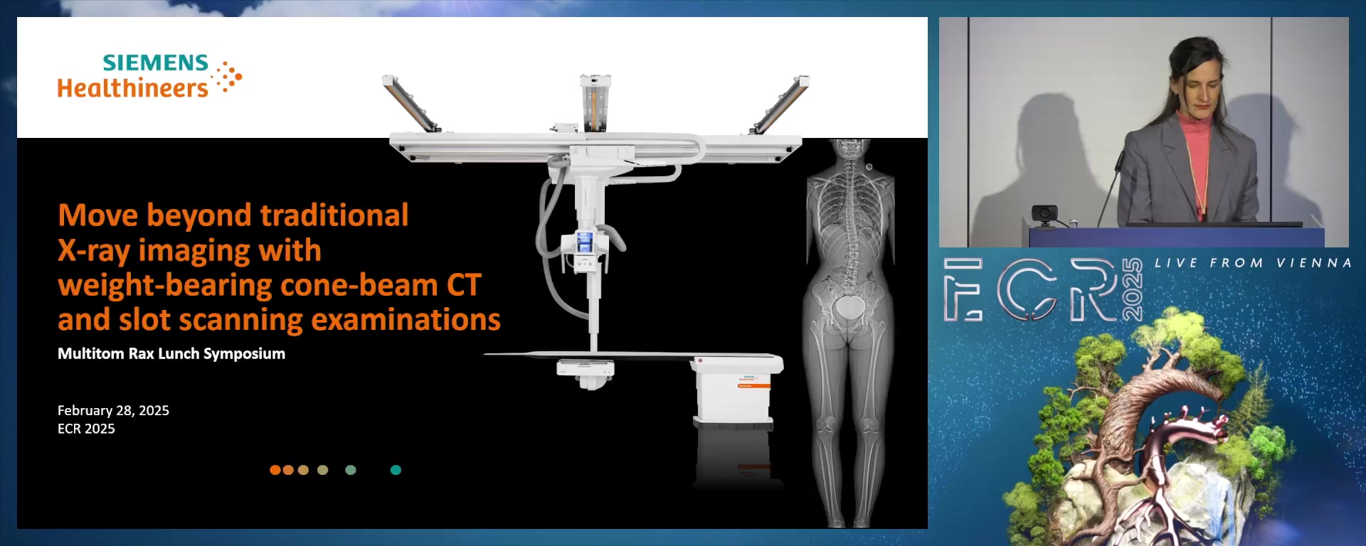
IND 18 - Move beyond traditional X-ray imaging with weight-bearing cone-beam CT and slot scanning examinations
Lectures
1
Move beyond traditional X-ray imaging with weight-bearing cone-beam CT and slot scanning examinations (Complete Session)
60:00Anna Rasche, Forchheim / DE, Arttu Peuna, Jyväskylä / FI, Marcus Treitl, Munich / DE, Sophia Freya Ulrike Blum, Dresden / DE, Manuel Scholl, Werneck / DE, Madeleine Hertel, Forchheim / DE
Musculoskeletal imaging has been part of the regular examination spectrum in radiology for over 100 years. However, precise measurements within these examinations require additional calibration on stitched imaging to avoid distortions. CT imaging may complement the examinations with their precise, superimposition-free images, but still lack the possibility to acquire functional weight-bearing images; and the relatively high CT radiation dose is only accepted.
With the advanced features Real3D and True2scale Body Scan, the Twin Robotic X-ray system Multitom Rax offers versatile solutions. Real3D offers three-dimensional X-ray examinations of the lower extremities and lumbar spine under physiological weight-bearing conditions which are relevant for numerous orthopedic applications. At the same time, True2scale Body Scan enables length-extended geometrically accurate, and dose reduced images in slot scanning technique, which can be used by orthopedists and surgeons for accurate surgical planning and therapy progress monitoring.
In this ECR Radiology Symposium, clinical experts from a wide range of disciplines will present new areas of application for Multitom Rax. Multidisciplinary clinical experts will:
• discuss the clinical possibilities with 3D weight-bearing examinations in a standing position
• demonstrate how important detailed and geometrically accurate imaging is for surgical planning
• show the possibilities for reducing radiation dose
5 min
Welcome and Introduction
Anna Rasche, Forchheim / Germany
- Discuss the clinical possibilities with 3D weight-bearing examinations in a standing position
- Demonstrate how important detailed and geometrically accurate imaging is for surgical planning
- Show the possibilities for reducing radiation dose
10 min
Effects of a multicomponent exercise regimen on subchondral bone, cartilage, and inflammation markers in postmenopausal women with knee osteoarthritis
Arttu Peuna, Jyväskylä / Finland
A randomized controlled study using weight-bearing CT examinations of the knee.
Recent advancements in cone-beam computed tomography (CBCT) have enabled detailed 3D imaging of the knee joint under load, offering better insights into joint behavior and allowing for precise evaluation of bone density, content, and joint space narrowing.
CBCT, combined with Convolutional Neural Network techniques, can automate knee osteoarthritis (OA) severity assessments using the Kellgren-Lawrence scale.
A randomized controlled study aims to explore the effects of a multicomponent exercise program, combining resistance and step-aerobic training, on subchondral bone, articular cartilage, inflammation, and metabolomic markers in postmenopausal women with knee OA.
The goal is to develop safe and effective exercise strategies that improve cartilage and bone health, prevent disability, and enhance mobility and quality of life for those affected by knee OA.
10 min
Geometric accuracy and radiation exposure on weight-bearing CT examinations of the lower limb
Marcus Treitl, Munich / Germany
CBCT and MSCT showed a high level of agreement in 3D imaging of the lower limb and assessing alignment and joint level metrics. By offering 3D imaging under physiological load, CBCT has the potential to improve preoperative planning in clinical use. A higher radiation dose must be addressed by further examinations with this model at even lower exposure doses.
10 min
Dose and image quality for whole-spine slot-scanning x-ray imaging compared with stitched radiography
Sophia Freya Ulrike Blum, Dresden / Germany
Repeated whole-spine imaging, especially in children and adolescents with scoliosis, causes significant cumulative radiation exposure. Low-dose slot-scanning imaging can reduce this exposure. This talk presents clinical experience and data from a University hospital, focusing on radiation exposure and image quality in these vulnerable patients.
10 min
Surgery planning with whole-leg slot-scanning x-ray imaging for accurate measurements of the leg-length
Manuel Scholl, Werneck / Germany
After implantation of an artificial hip joint, the resulting leg length is an important outcome for the patient. The standard method to determine the leg length is the preoperative pelvic overview x-ray images, in which the measurements for the surgery are performed and the outcome estimated. As this image view does not include the complete leg, estimations of postoperative leg length might vary. With slot-scanning x-ray imaging, precise weight-bearing images of the whole leg including the pelvis can be acquired and offer a more accurate and does-reduced estimation of the resulting leg length.
15 min
Q&A and Discussion
Madeleine Hertel, Forchheim / Germany