Refresher Course: Neuro
RC 111 - Post-neurosurgery: what to expect on imaging
5 min
Chairperson's introduction
Sofie Van Cauter, Holsbeek / Belgium
15 min
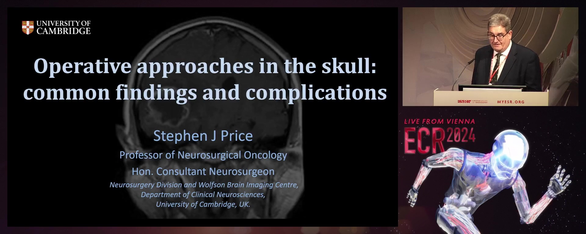
Operative approaches in the skull: common findings and complications
Stephen Price, Cambridge / United Kingdom
Learning Objectives
1. To name three routes of CSF leakage post-operatively.
2. To describe the concept of 'brain shift' using image guidance.
3. To list three potential causes of a post-operative subdural collection.
4. To describe why contrast-enhancing tumours may be left behind following surgery.
2. To describe the concept of 'brain shift' using image guidance.
3. To list three potential causes of a post-operative subdural collection.
4. To describe why contrast-enhancing tumours may be left behind following surgery.
15 min
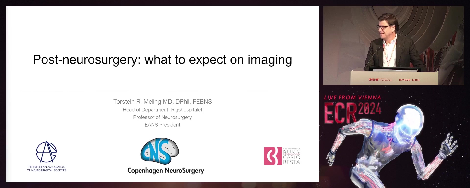
Post-neurosurgery artifacts on MRI imaging
Torstein Ragnar R. Meling, Copenhagen / Denmark
Learning Objectives
1. To illustrate imaging features and artefacts caused by hemostatic agents in cranial surgery.
2. To explain the CT and MRI aspect of duraplasty materials and bone flap fixation devices.
3. To identify how to recognise imaging complications resulting from implanted materials.
2. To explain the CT and MRI aspect of duraplasty materials and bone flap fixation devices.
3. To identify how to recognise imaging complications resulting from implanted materials.
15 min
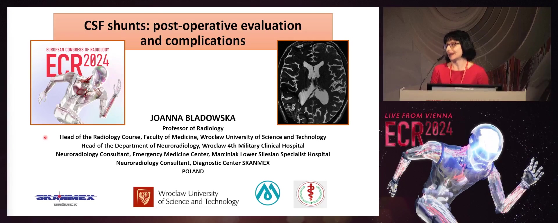
CSF shunts: post-operative evaluation and complications
Joanna Bladowska, Wroclaw / Poland
Learning Objectives
1. To define the most common types of CSF shunts as well as the application of different imaging methods for the evaluation of shunt malfunction.
2. To list and describe the most common complications (including mechanical failure, infection, ventricular loculation, overdrainage and the specific ones related to the shunt type) and discuss the key findings that may be useful for the correct diagnosis.
3. To be able to identify possible outliers and pitfalls on imaging.
10 min
Panel discussion: How to improve post-operative reporting in neuroradiology
