Research Presentation Session: Radiographers
RPS 314 - Research evidence to inform and shape the future
Lectures
1
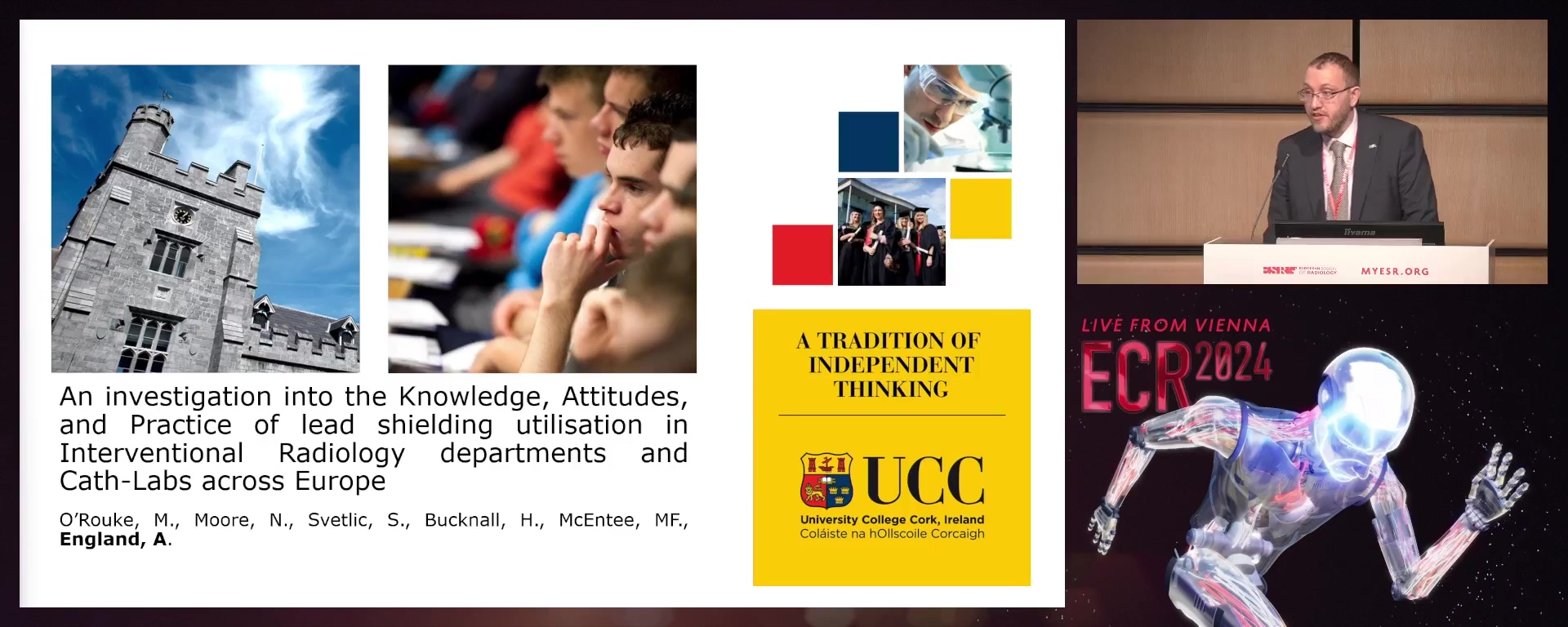
An investigation into the knowledge, attitudes, and practice of lead shielding utilisation in interventional radiology departments and cath-labs across Europe
07:00Andrew England, Cork / IE
2
Review of diagnostic reference levels (DRLs) in interventional neuroradiology (INR)
07:00Marvin Grech, Ghaxaq / MT
3
Public awareness and preferences for medical radiation risk information
07:00Gráinne Alison Curran, Cork / IE
4
Correlating breast lesion dimensions: imaging vs pathological results
07:00Laura Martins Segura de Jesus, Vila Real de Santo António / PT
5
Patients’ experience to MRI examinations: a systematic qualitative review with metasynthesis
07:00Isabel Nieto Alvarez, Waldkirch / DE
6
Reporting radiographers in Europe: an online survey
07:00Malene Roland Vils Pedersen, Vejle / DK
7
Density bone evaluation by ECHOS: a pilot study
07:00Rute Santos, Coimbra / PT
8
Liver fibrosis and steatosis ultrasound screening: a FibroScan study
07:00Rute Santos, Coimbra / PT
RPS 314-3
7 min
An investigation into the knowledge, attitudes, and practice of lead shielding utilisation in interventional radiology departments and cath-labs across Europe
Andrew England, Cork / Ireland
Author Block: M. O'Rouke1, N. Moore1, S. Svetlic2, H. L. Bucknall3, M. F. F. McEntee1, A. England1; 1Cork/IE, 2Milan/IT, 3London/UK
Purpose: According to current literature, there is a lack in the knowledge, attitudes and practices (KAP) of radiation protection (RP) among interventional radiology (IR) and Cath-lab staff. To the best of the authors’ knowledge, there are no studies investigating the radiation protection KAP of radiology staff within IR departments and cath-labs across Europe. This study aims to determine the RP KAP of staff within IR and cath-labs across Europe and the associated influencing factors.
Methods or Background: A cross-sectional study in the form of an online questionnaire was developed. Participation was advertised online via online platforms and through email. Inclusion criteria included qualified healthcare professionals currently working in Interventional Radiology and cath-labs across Europe. Section 1 of the questionnaire consisted of questions regarding demographic data. Section 2 comprised of questions regarding RP training and protocols. Section 3 involved questions regarding the use of different types of RP lead shields, both personal and co-worker use. Section 4 concluded the questionnaire with questions regarding other methods of minimising radiation dose within the departments.
Results or Findings: A total of 178 responses to the questionnaire were recorded. Most respondents were female (72%), radiographers (75%) and within the age bracket of 25-34 (46%). Only (53%) had ever received RP specific training and the majority (63%, 80) of respondents were currently practicing in Ireland.
Conclusion: The KAP of IR and Cath-Lab staff regarding RP within departments across Europe is low. The unavailability of basic radiation protection tools and RP specific training courses/modules were some of the reasons for sub-optimal self-protection against ionising radiation exposure among the respondents. To avoid unnecessary exposure to themselves, co-workers, and patients, it is suggested that medical professionals be equipped with appropriate training and RP tools.
Limitations: Self-reporting questionnaire limits this study.
Funding for this study: No funding was obtained for this study.
Has your study been approved by an ethics committee? Yes
Ethics committee - additional information: This study was ethically approved by the Medical School SREC - University College Cork.
RPS 314-4
7 min
Review of diagnostic reference levels (DRLs) in interventional neuroradiology (INR)
Marvin Grech, Ghaxaq / Malta
- Author Block: M. Grech, F. Zarb, R. Grech, P. Bezzina; Msida/MTPurpose: The objective of this study was to evaluate radiation dose quantities and methodology for establishing diagnostic reference levels DRLs (3rd quartile/75th percentile) for interventional neuroradiology (INR).Methods or Background: Four different databases: Scopus, Web of Science, PubMed, and Pro Quest were utilized using four keywords: Air Kerma-Area Product (PKA), Fluoroscopy Time (FT), Reference Air Kerma (CAK) and DRLs and their synonyms to search literature in English within the last decade.Results or Findings: DRLs were recorded from 38 studies for different Neurointerventional procedures extracted from the literature for PKA (Gy/cm2), FT (Minutes), and RAK (mGy). Procedure - Number of studies / PKA (Gy/cm2) Mean (min/max) / FT (minutes) Mean (min/max) / RAK (mGy) Mean (min/max), Cerebral Angiography - 29 /
- 64 (41/256.65) / 11.73 (6/28.4) / 657.74 (289/921.1), Stroke Thrombectomy - 8 / 163.57 (110/225.1) / 41.34 (30/44/7) / 1012.56 / (730/1590), Aneurysm Coiling - 27 / 254.06 (52.1/487.4) / 54.51 (16/90) / 3309.89 (505/4750) AVM/AVF Embolisation - 9 /
- 63 (206.4/550) / 86.37 (58.57/535) / 4130.20 (2350/6000). Statistically significant (p<
- 05) variations are evident between procedures based on their complexity. Other variations based on equipment type, operator experience and methods of data collection were also noted. The authors also sought to identify potential reasons for such discrepancies within the included studies. Conclusion: To date, few studies have published data regarding DRL’s in INR procedures. The evident variation in DRL quantities, warrants the need for optimization strategies, and guidelines for quality control and to reduce procedural radiation doses as per the “ALARA” principle. Establishment and analysis of DRLs in INR procedures is essential for subsequent optimisation of radiation doses in INR procedures.Limitations: This is a literature review and is based on the information available within the reviewed papers.Funding for this study: No funding was received for this study.Has your study been approved by an ethics committee? Not applicableEthics committee - additional information: Not applicable for this study.
RPS 314-5
7 min
Public awareness and preferences for medical radiation risk information
Gráinne Alison Curran, Cork / Ireland
- Author Block: N. Mernagh, A. England, N. Moore, R. Young, M. F. F. McEntee; Cork/IEPurpose: New legislation says that healthcare workers must inform patients of the radiation dose and risks associated with medical imaging. Literature suggests the public are being poorly informed. There has been little research carried out where comparisons of risk have been evaluated in terms of patient understanding. The aim of this study is to investigate radiation awareness and preferences the public may have on radiation risk communication prior to medical imaging examinations.Methods or Background: A cross-sectional study was used to obtain quantitative and qualitative data from the public. The online questionnaire was designed using Qualitrics XM. The target participants were members of the public over 18 years, irrespective of whether they have undergone previous imaging. Data was analysed using Microsoft Excel.Results or Findings: A total of 413 participants completed the survey. Majority (n=364,
- 1%) had previously had imaging. Most respondents rated their knowledge on radiation risks as poor (n=213/401, 53.1%). Participants were asked who should be communicating these risks with them, the person carrying out the examination (usually the radiographer) received the most responses (n=334, 34.7%), while the referrer was the second most common (n=224, 23.3%). For the risk communication methods, majority stated very helpful for the associated risk is negligible/minimal/very-low/low/moderate (34.2%), followed by the associated risk of cancer is 1 in 2000 (33.4%). Conclusion: The public have a poor awareness of radiation risks from medical imaging. Multiple responses to who should communicate risk suggests a multidisciplinary approach is warranted. Simple language to convey risk is preferred along with phrases which directly address cancer risk.Limitations: Self-reporting survey limits the scope of this study.Funding for this study: No funding was received for this study.Has your study been approved by an ethics committee? YesEthics committee - additional information: Medical School SREC - University College Cork approved this study.
RPS 314-6
7 min
Correlating breast lesion dimensions: imaging vs pathological results
Laura Martins Segura de Jesus, Vila Real de Santo António / Portugal
- Author Block: L. M. S. d. Jesus, A. F. C. L. Abrantes, S. I. Rodrigues, L. F. Carvalho, L. P. V. Ribeiro; Faro/PTPurpose: This study aims to establish the correlation between breast neoplasm dimensions as measured via Mammography (MGM), Ultrasonography (US), and Magnetic Resonance Imaging (MRI), and the real tumour dimensions obtained from anatomopathological reports of surgical specimens.Methods or Background: A retrospective study was carried out on 56 women diagnosed with breast cancer, between January 2021 and November 2022, in a Portuguese Public Hospital. The study assessed the size of the primary tumour using MGM, US, and breast MRI. Subsequently, we compared these measurements with those obtained from the anatomopathological examination.Results or Findings: Our findings revealed that, in MGM, US, and MRI, there was an overestimation in 34/56 (
- 7%), 18/56 (32%), and 31/56 (55%) cases, respectively, and an underestimation in 18/56 (32%), 34/56 (60.7%), and 19/56 (33%) cases, respectively. We observed an absolute agreement in measurements for 4/56 cases between MGM and US when compared to pathological anatomy, and 6/56 cases between MRI and pathological anatomy. Conclusion: Our study concluded that there were no significant differences in tumour size measurements between MGM and MRI when compared to pathological anatomy. Additionally, we found that factors such as histological type or breast density did not significantly influence measurement accuracy. Notably, MRI exhibited the highest accuracy in evaluating lesion dimensions, demonstrating a superior correlation with tumour measurements obtained through anatomopathological examination.Limitations: The main limitations of this study are the small sample size due to the number of patients who underwent neoadjuvant chemotherapy, the lack of measurements of all the lesions in all the available imaging modalities due to the external origin of the patients, and the issue of measurement error.Funding for this study: Not applicable for this study.Has your study been approved by an ethics committee? YesEthics committee - additional information: This study was funded by the Uaif: 159-
RPS 314-7
7 min
Patients’ experience to MRI examinations: a systematic qualitative review with metasynthesis
Isabel Nieto Alvarez, Waldkirch / Germany
Author Block: I. Nieto Alvarez1, J. Madl1, L. Becker1, O. Amft2; 1Erlangen/DE, 2Freiburg/DE
Purpose: The review informs practitioners, patients, and policymakers of observations for future research, and describes the experience and characterising factors in MRI through the adult patients’ voice for guidance of future efforts addressing patient needs.
Methods or Background: Patients often mention distress, anxiety or claustrophobia related to MRI, resulting in no-shows, disturbances of the workflow, and lasting psychological effects. Patients’ experience varies and is moderated by socio-demographic aspects alongside the clinical condition. While qualitative studies help understand individuals’ experiences, to date a systematic review and aggregation of MRI individuals’ experience is lacking.
We conducted a systematic search in PubMed, Scopus, Web of Science, and PsycInfo databases according to the PRISMA guidelines to identify primary studies reporting patients’ responses to MRI. For quality appraisal, the Joanna Briggs Institute (JBI) tools were used. Metasynthesis, a concept map, and meta-aggregation were used for data synthesis.
Results or Findings: We identified eight papers on patients’ experience description for qualitative meta-summary (294 fulltexts, 46 studies with sufficient quality, 49 quantitative studies), 220 patients in total. Meta-aggregation of 144 patient quotes answered: (1) experiences before, at the scanning table, during, and after an MRI, (2) differences based on clinical condition, and (3) characterising factors, including coping strategies, look-and-feel of medical technology, interaction with professionals, and information. Noteworthy across studies was the difficulty with the confined space, fear of results, need for information and coping strategies.
Conclusion: Our findings provide a foundational description of adult patients’ MRI experience, revealing themes and characterising factors at each stage of the procedure through the patients' voice.
Limitations: Most publications lack participants' health literacy level, occupation, developmental conditions, ethnicity, or country of origin. Studies were mostly conducted in university hospitals. Interviews’ raw data unavailability impeded computer-aided analysis.
Funding for this study: This research did not receive any specific grant from funding agencies in the public, commercial, or not-for-profit sectors.
Has your study been approved by an ethics committee? Not applicable
Ethics committee - additional information: The study was conducted according to a systmatic literature review with meta-summary and metasynthesis of published patient quotes.
RPS 314-8
7 min
Reporting radiographers in Europe: an online survey
Malene Roland Vils Pedersen, Vejle / Denmark
- Author Block: M. R. V. Pedersen1, J. Jensen2, C. Senior3, N. Gale4, C. J. Heales4, N. Woznitza5; 1Vejle/DK, 2Odesa/DK, 3Axminster/UK, 4Exeter/UK, 5London/UKPurpose: Reporting radiographers undertake an important role in health care and for the radiographer profession ingeneral. First introduced in the UK, and now in several other European countries. The objective was to investigate the workforce of reporting radiographers across the European Federation of Radiographer Societies (EFRS) community.Methods or Background: A voluntary anonymous 34-item electronic survey was distributed online using social media accounts such asTwitter, Facebook, and LinkedIn cover a wide range of topics relating to professional roles, advanced practice, education, and seniority. The questionnaire was distributed during a 12-week period in Results or Findings: A total of 345 individual responses were received from 15 countries with majorities of respondents fromUnited Kingdom (n=245, 71%) and Denmark (n=66, 19%). The mean age was
- 9 (S.D 9.8), similar for females, 42.5 (S.D 9.0), and men 40.9 years (S.D 9.7). Most reporting radiographers worked in public hospitals (90%). The vast majority of the respondents (n=270, n=94%) authored and signed their own clinical reports while a minority (n=18, 6%) stated that their reports were checked by radiologists. Conclusion: The survey highlights the scope of practice of reporting radiographers working in Europe. Reporting is becoming a career path for an increasing number of radiographers across Europe.Limitations: A limitation is that the survey was published in English, so language may have formed a barrier toparticipation within non-English speaking countries.Funding for this study: No funding was received for this study.Has your study been approved by an ethics committee? YesEthics committee - additional information: Research Ethics Committee of the University of Southern Denmark approved this study.
RPS 314-9
7 min
Density bone evaluation by ECHOS: a pilot study
Rute Santos, Coimbra / Portugal
- Author Block: R. Santos, J. Nobrega; Coimbra/PTPurpose: The purpose of this study was to correlate the BMD values obtained through ECHOS with the individual characteristics of the participants and to compare the BMD values (T-Score and Z-Score) between DEXA and ECHOS.Methods or Background: Two hundred and nineteen participants were submitted to an ECHOS evaluation and 16 of 219 participants were evaluated by DEXA. All of the participants were evaluated on lumbar spine. All participants answered a sociodemographic questionnaire with factors influencing bone mineral density (eating habits, smoking, physical activity) and signed informed consent.Results or Findings: Of the 219 participants, 68% were female,
- 8% practiced physical activity, 5.9% had hormonal pathologies, 53% were between 22 and 59 years and 11.4% smoked regularly in, however, after comparing the BMD there was a significant correlation with weight, consumption of tobacco and foods rich in calcium but not with the other variables this in the first study, however the second study revealed the existence of a significant correlation between the values of Z-Score but despite this in the values of T-Score was not verified. Conclusion: In short, this study showed that some factors such as smoking, weight and calcium intake may influence the values obtained in ECHOS and also the existence of agreement between the two techniques used to assess BMD. ECHOS, having the advantages of ultrasound, can be an imaging method used to screen for osteoporosis or osteopenia. However future studies must be developed.Limitations: Some questionnaires were not completed by participants, the equipments were not always available, not allowing the evaluation of same number of subjects.Funding for this study: Not applicable for this study.Has your study been approved by an ethics committee? YesEthics committee - additional information: The study in question obtained authorisation from the ethics of the Polytechnic Institute of Coimbra (IPC) with number 161_CEIPC_
RPS 314-10
7 min
Liver fibrosis and steatosis ultrasound screening: a FibroScan study
Rute Santos, Coimbra / Portugal
- Author Block: R. Santos, B. Fonseca; Coimbra/PTPurpose: The aim of this study was to correlate the values of hepatic fibrosis and steatosis obtained through FibroScan with the individual characteristics of the participants, aiming to identify risk factors associated with the presence or absence of these conditions.Methods or Background: Data collection was conducted through a screening initiative. The exclusion criteria were used: people weighing more than 30 kg/m2, people under 18 years of age and participants who have already undergone kidney or liver transplants. The population was invited to participate in the evaluation, and the entire procedure was explained to them. Volunteers underwent a FibroScan examination and answered sociodemographic questions.Results or Findings: Out of 169 participants, 29% were men,
- 5% were overweight and obese, 15.9% had high cholesterol, 2.9% had diabetes, and 13% had hypertension (HTN). After analysis, 92 participants were selected for fibrosis evaluation and 107 for hepatic steatosis evaluation. The majority of participants did not have fibrosis or hepatic steatosis (76.1% and 64.4%, respectively). 7.6% of participants had moderate to severe fibrosis, and 3.3% had cirrhosis. Regarding hepatic steatosis, 6.6% had the more severe grades. Hepatic fibrosis was not associated with diabetes, cholesterol, HTN, sex, or age. Hepatic steatosis was associated with diabetes, cholesterol, HTN, and age but was not related to the participants' sex. Conclusion: Hepatic fibrosis and steatosis may be related to risk factors. It is important to screen the population so that an early diagnosis of liver disease can be made and possible worsening and complications can be avoided.Fibroscan is a technique that allows a rapid assessment of the liver and its changes, making it ideal for broad population screening.Limitations: Some of the participants did not answer all the questions. The fibroscan examination was performed by more than two radiographers.Funding for this study: Not applicable for this study.Has your study been approved by an ethics committee? Not applicableEthics committee - additional information: It was a student initiative, where only the data was used afterwards, so no ethical approval was sought.