Research Presentation Sessions: Radiographers
RPS 2014 - Risk reduction and quality assurance in radiographic practice
Lectures
1
Introduction by the moderator
02:00Luis J.O.C. Lança, Lisbon / PT, Annalisa Trianni, Trento / IT
2
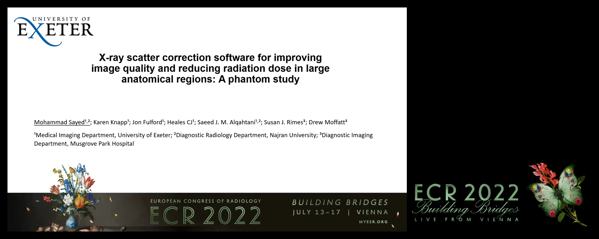
X-ray scatter correction software for improving image quality and reducing radiation dose in large anatomical regions
08:00Mohammad Sayed, Exeter / UK
3
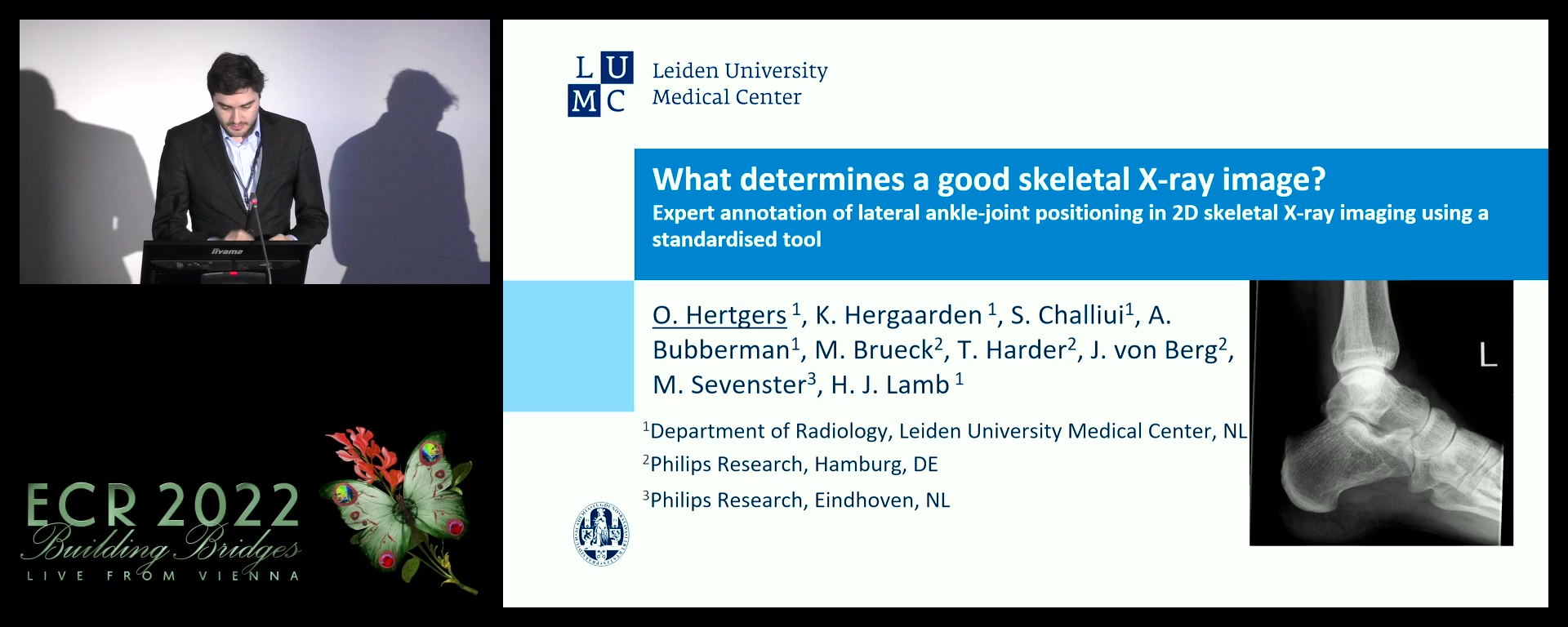
What determines a good skeletal X-ray image? Expert annotation of lateral ankle-joint positioning in 2D skeletal X-ray imaging using a standardised tool
08:00Omar Hertgers, Den Haag / NL
4

Trabecular bone score (TBS) role in fracture risk assessment
08:00Liliana Rodrigues, Exeter / UK
5
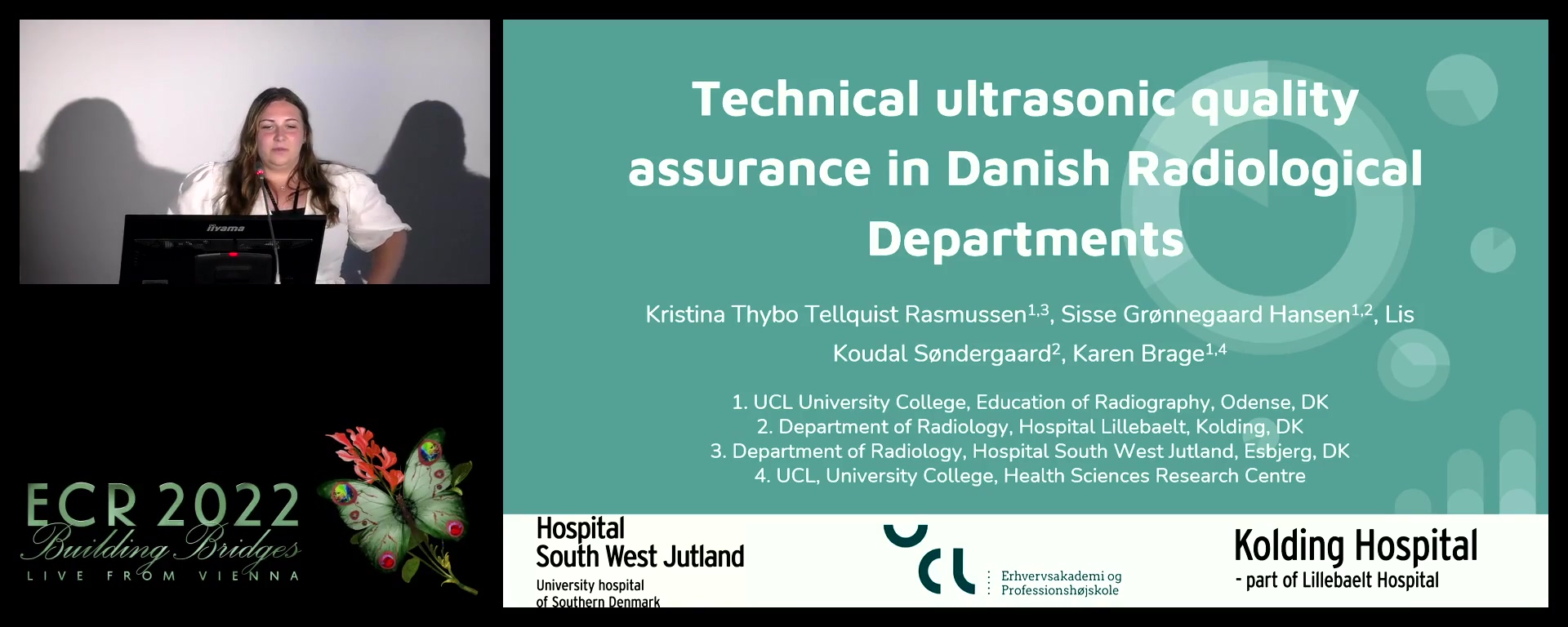
Technical ultrasonic quality assurance in Danish radiological departments
08:00Kristina Rasmussen, Børkop / DK
6
Image quality evaluation of on-centre and off-centre FOV in cardiac CT examination (spatial resolution and motion artifacts)
08:00Katsumi Tsujioka, Toyota-City Aichi / JP
7

Dual-energy CT: reduction of metal artifacts in the skeletal-muscle system
08:00Mário Monteiro, COIMBRA / PT
8
Characterisation of national radiotherapy departments: organisation, occupational exposure values and local diagnostic reference levels for breast and prostate CT-planning
08:00Rafaela Guisantes, Coimbra / PT
9
Development of a patient face recognition system in the radiology department (demonstration study of a face mask-enabled face recognition system in a CT examination environment)
08:00Hiroyuki Ota, Kashiwa,Chiba pref / JP
10
Explaining radiation risk: an investigation of approaches preferred by Irish patients
08:00Evan Comiskey, Dublin / IE
11
NiFi: an open source application for secure and efficent handling of image data for radiographic studies
08:00Martin Kusk, Esbjerg / DK