Refresher Course: Head and Neck
RC 1908 - Skull base imaging
Lectures
1
Chairperson's introduction
--:--Timothy Beale, London / UK
2
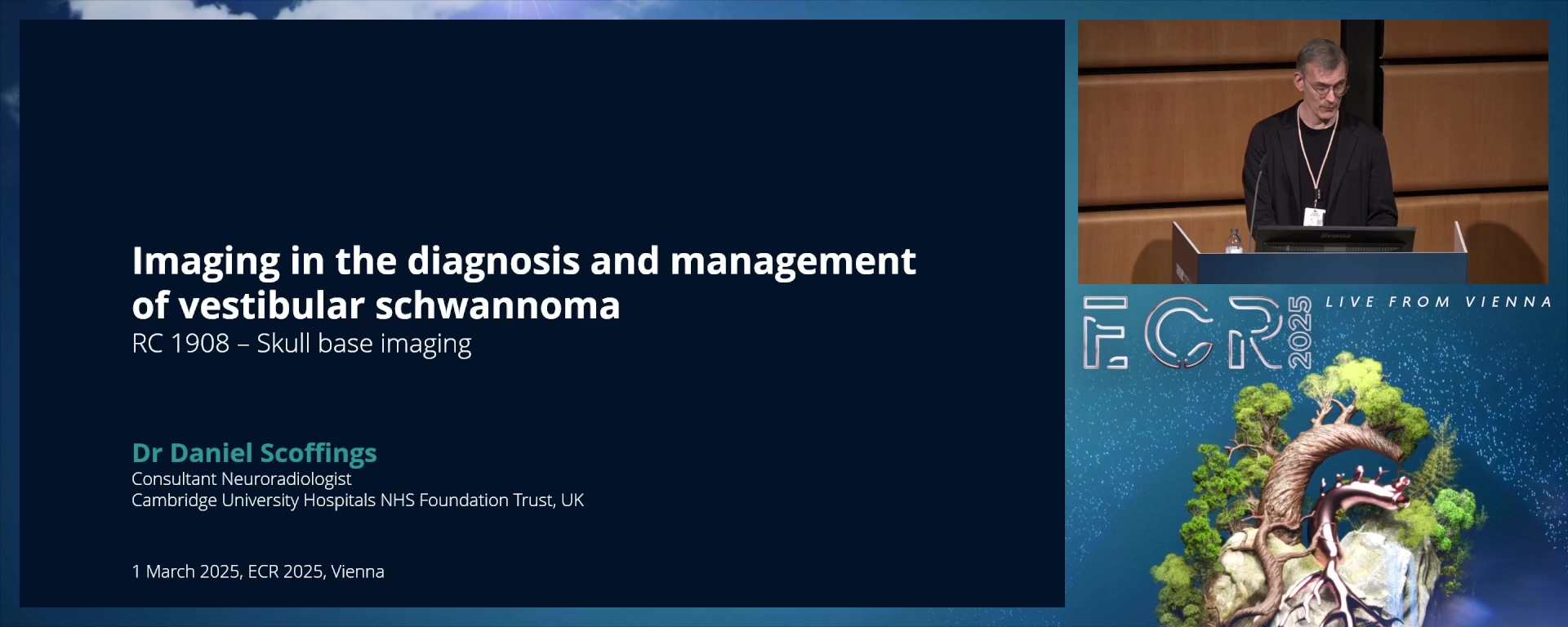
Imaging in the diagnosis and management of the vestibular schwannoma
15:00Daniel Scoffings, Cambridge / UK
3
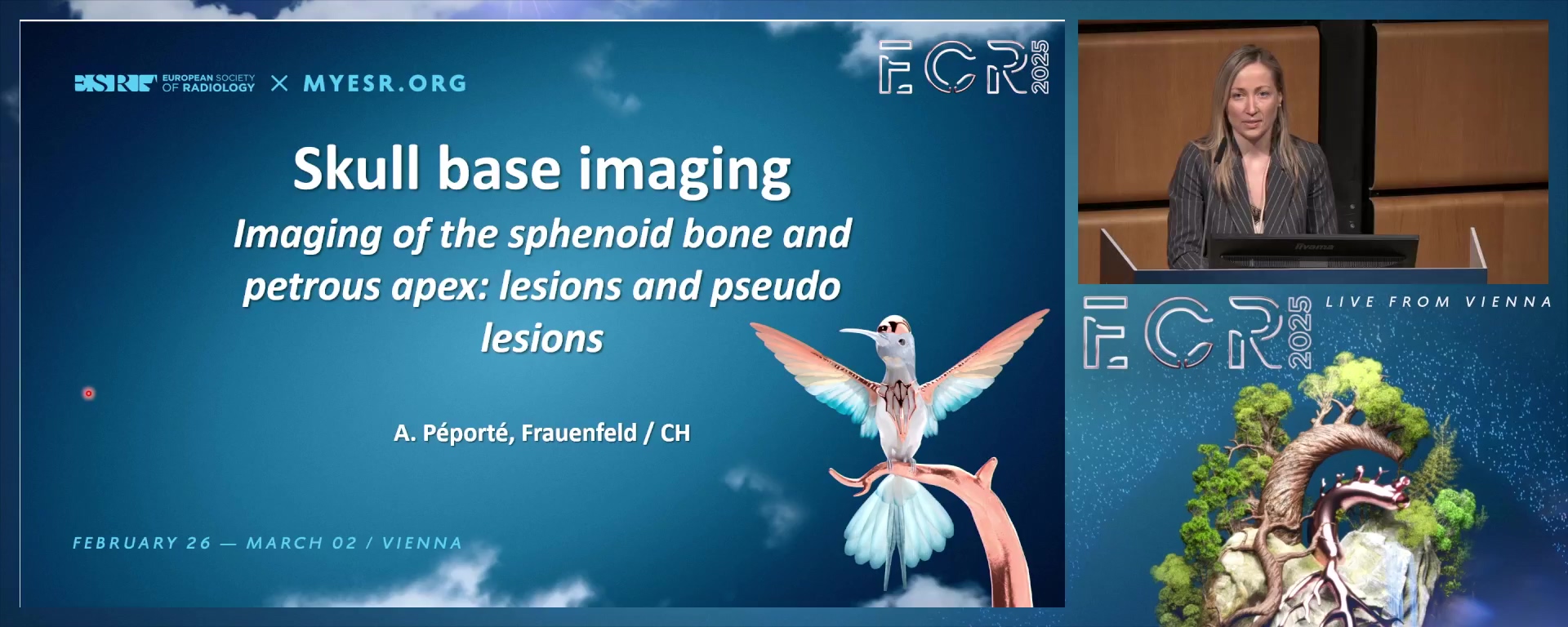
Imaging of the sphenoid bone and petrous apex: lesions and pseudo lesions
15:00Anne Renée Juliette Péporté, Frauenfeld / CH
4
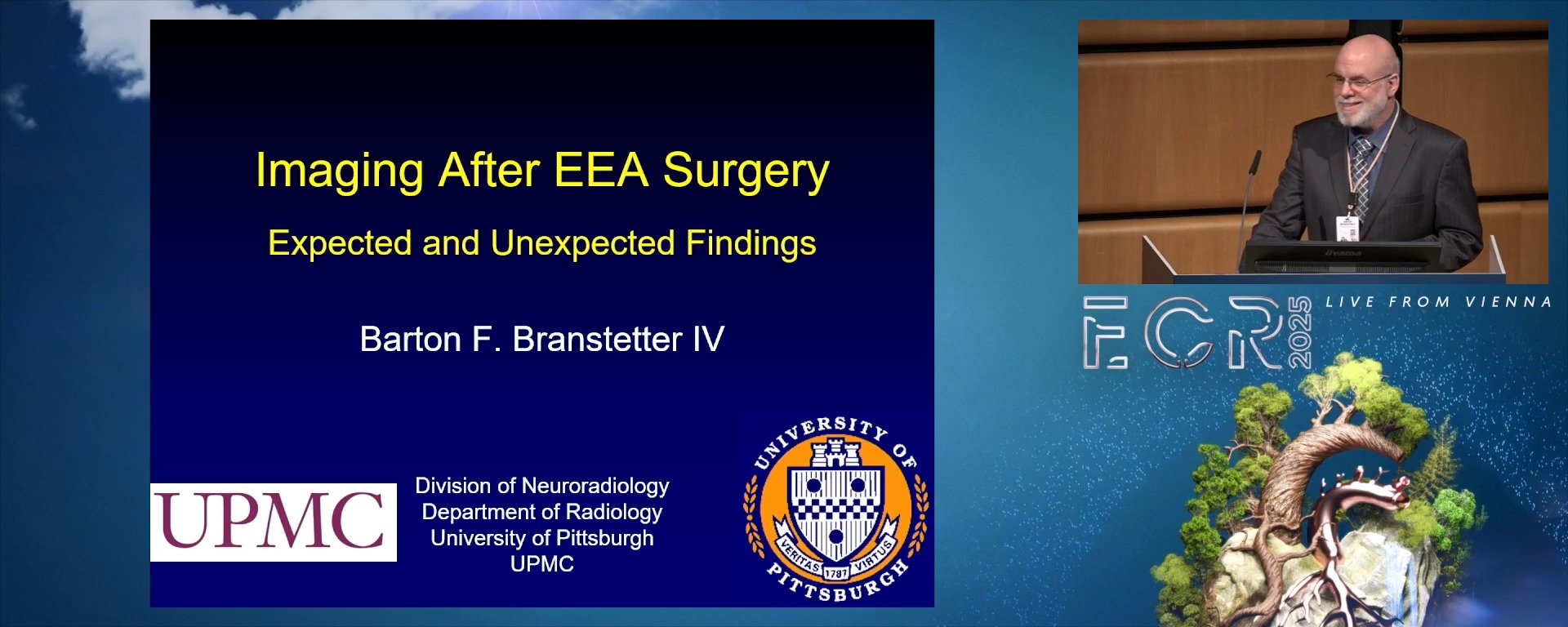
Imaging after expanded endonasal skull base surgery: expected and unexpected findings
15:00Barton F. Branstetter IV, Wexford / US
5
Panel discussion: Tips and pitfalls in skull base imaging. What I wish someone had told me
10:00Panel discussion: Tips and pitfalls in skull base imaging. What I wish someone had told me
5 min
Chairperson's introduction
Timothy Beale, London / United Kingdom
15 min
Imaging in the diagnosis and management of the vestibular schwannoma
Daniel Scoffings, Cambridge / United Kingdom
- To identify the common and rare imaging findings in vestibular schwannoma and to confidently differentiate them from its most common mimics.
- To outline the current therapeutic strategies and to discuss the most important imaging features which impact management decisions.
- To describe the expected post-treatment appearances and indicate the features suggestive of tumour recurrence during follow-up.
15 min
Imaging of the sphenoid bone and petrous apex: lesions and pseudo lesions
Anne Renée Juliette Péporté, Frauenfeld / Switzerland
- To recognise the most common "don't touch" lesions, differentiate them from malignant lesions and describe their implications according to their complex anatomical connections.
- To narrow the differential diagnosis by defining the behaviour and origin of the lesion as well as other imaging characteristics.
- To apply a feasible algorithm for the diagnostic and therapeutic management of such lesions in clinical practice.
15 min
Imaging after expanded endonasal skull base surgery: expected and unexpected findings
Barton F. Branstetter IV, Wexford / United States
- To understand the different types of flap reconstructions used in endoscopic endonasal skull base reconstructions. 2.To integrate a diagnostic approach for differentiating treatment change from residual disease on follow-up imaging.3.To differentiate the expected postsurgical imaging appearance from that of surgical complications.
10 min
Panel discussion: Tips and pitfalls in skull base imaging. What I wish someone had told me