E³ - Young ECR Programme: Students Session
S 10 - Students session 1
Lectures
1
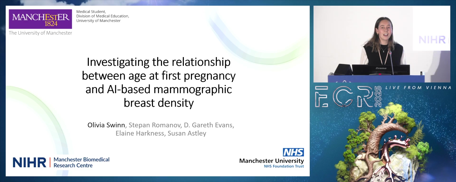
Assessing the relationship between AI-based mammographic breast density and age at first pregnancy
08:00Olivia Swinn, Manchester / UK
2
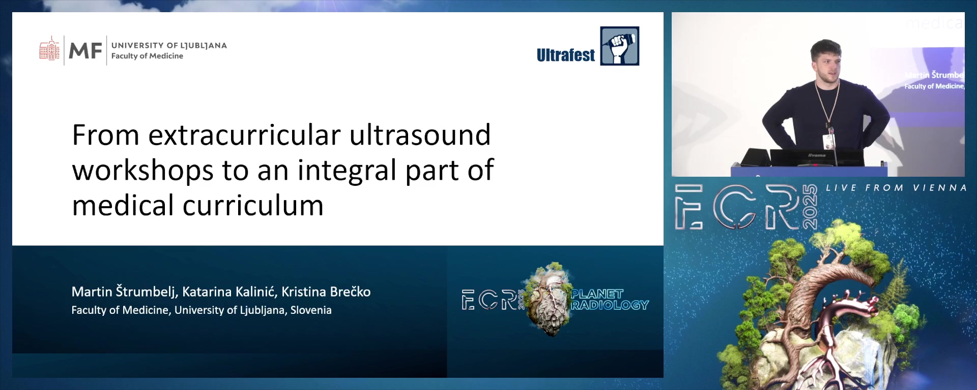
From extracurricular ultrasound workshops to an integral part of medical curriculum
08:00Martin Štrumbelj, Ljubljana / SI
3
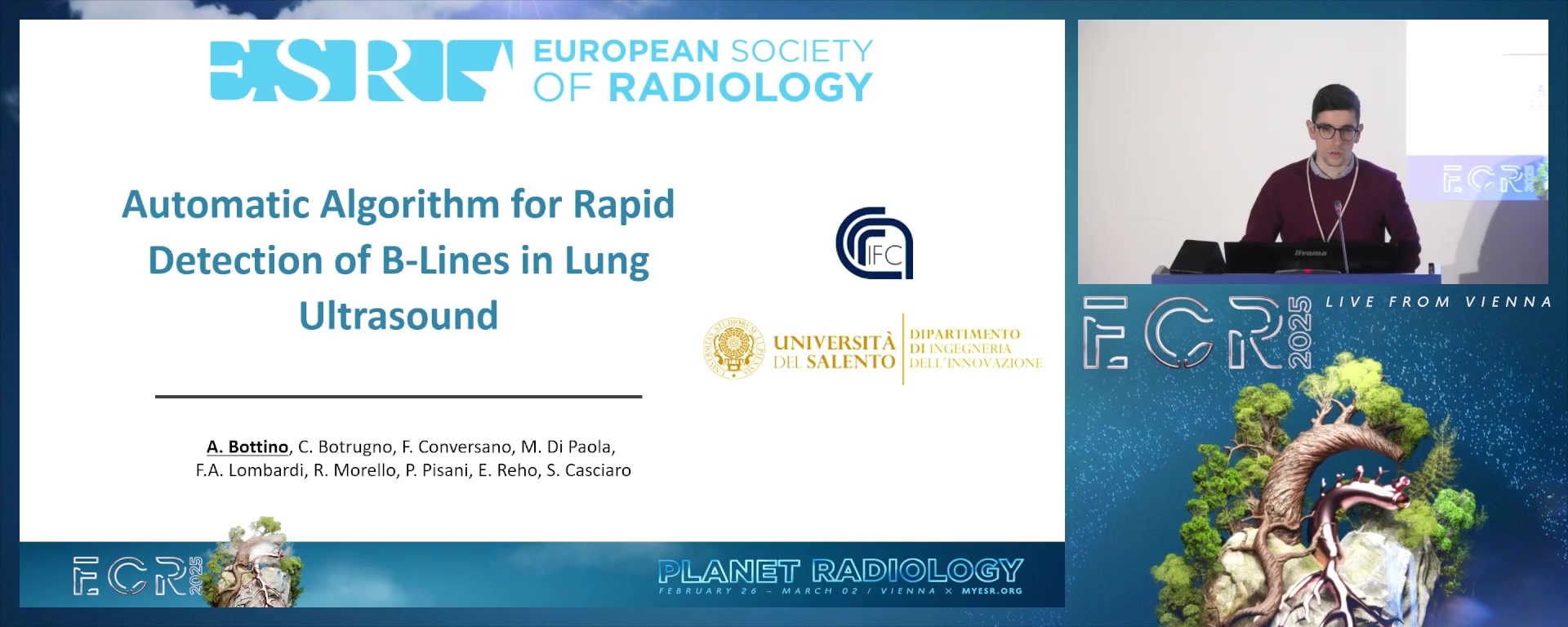
Automatic Algorithm for Rapid Detection of B-Lines in Lung Ultrasound
08:00Alberto Bottino, Lecce / IT
4
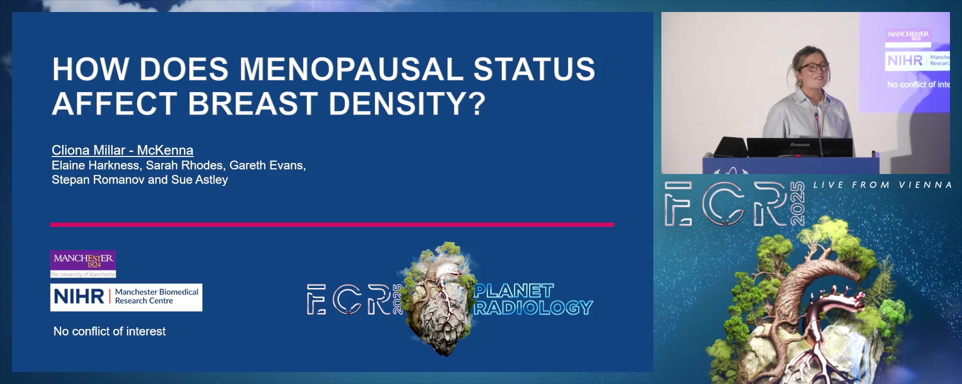
Menopausal status and breast density
08:00Cliona Mckenna, White - Irish / UK
5
Effects of gender-affirming hormone therapy (GAHT) in transgender individuals assigned male at birth (AMAB): evaluation of breast development and radiological characteristics
08:00Alice Canetti, Sanremo / IT
6
Comparative Analysis of Medical Students' Perspectives on Radiology: Pre- and Post-Pandemic Insights
08:00Cristiana Marcu, Bucharest / RO
7
Combined use of MRI-derived maps of fractional tumor burden and apparent diffusion coefficient to identify tumor progression in high-grade astrocytoma patients
08:00Siem Herings, Nijmegen / NL
8
Can student participation in a radiological anatomy Peer-Assisted Learning program (PALP) impact academic life?
08:00João Vítor Leal Souza, Salvador / BR
9
Artificial Intelligence: Pathway or Setback for Sustainable Radiology?
08:00Anca-Mihaela Necula, Craiova / RO
8 min
Assessing the relationship between AI-based mammographic breast density and age at first pregnancy
Olivia Swinn, Manchester / United Kingdom
Author Block: O. Swinn, S. Romanov, D. G. Evans, E. F. Harkness, S. M. Astley; Manchester/UK
Purpose: The aim of this study is to investigate the relationship between breast density and parity, assessed using the age at first pregnancy and an AI generated measure of breast density (MAI_VAS).
Methods or Background: This study uses a subset of the PROCAS study, a large-scale study that collected breast cancer risk information for over 40,000 women across Greater Manchester. Mean breast density was compared between women of different ages, ethnic origins, and different ages at first pregnancy. Statistical analysis was carried out using ANCOVA tests to investigate how breast density is influenced by parity. Initially univariate analysis was performed, followed by multivariate analyses considering age at consent to study, BMI, menopausal status, and ethnic origin.
Results or Findings: Nulliparous women and those with an age at first pregnancy over 35 demonstrated the highest mean breast densities at 34.1 and 35.6 respectively. Women who were youngest at the time of first pregnancy (under 25) had the lowest mean breast density (26.4), which was statistically significantly different from all other parity groups. There was no significant difference between the breast densities of women aged 30 to 34 and over 35 at the time of first pregnancy, and adjusting for ethnic origin made no statistical difference.
Conclusion: In conclusion, a positive correlation was observed between a higher age at first pregnancy, nulliparity and mammographic breast density.
Limitations: A limitation of this study could be that the demographics of the PROCAS participants and therefore this study cohort may not be entirely representative of both the screening and general population since uptake to PROCAS was relatively low and attendance at screening is not equal across all ethnic groups.
Funding for this study: Prof Sue Astley Prof D Gareth Evans are supported by the National Institute for Health and Care Research (NIHR) Manchester Biomedical Research Centre (BRC) (NIHR203308)
We would also like to thank the many radiographers in the screening programme and the PROCAS study staff for patient recruitment and data collection. PROCAS study was funded by the National Institute for Health Research (Reference Number RP-PG-0707-10031), with additional support from the Prevent Breast Cancer. This abstract presents independent research; views expressed are those of the authors and not necessarily those of the NHS, the NIHR or the Department of Health.
Has your study been approved by an ethics committee? Yes
Ethics committee - additional information: Ethics approval for the study was through the North Manchester Research Ethics Committee (09/H1008/81). Informed consent was obtained from all participants on entry to the PROCAS study.
8 min
From extracurricular ultrasound workshops to an integral part of medical curriculum
Martin Štrumbelj, Ljubljana / Slovenia
Author Block: M. Štrumbelj, K. Kalinić, K. Brečko; Ljubljana/SI
Purpose: Ultrafest was established in 2016 as a student project following rising demand from medical students lacking ultrasound training in their medical education. After years of organising hands-on point of care ultrasound (POCUS) workshops, our activities have been integrated in the newly adapted curriculum.
Methods or Background: Ultrafest is designed as a one-day event with lectures and hands-on POCUS workshops, performed by clinical specialists. To prepare, the students are given literature, a textbook written by mentors and students of the project in 2021. After the event, students complete an evaluation form, asking them to rate their confidence in performing POCUS examination before and after the workshops on a scale from one (poor skills) to five (excellent skills).
Results or Findings: Ultrafest was, over the seven years of workshops, attended by 620 students. The average self-assessment score nearly doubled from 2.0 before the workshops to 3.8 after, showing a significant improvement in the confidence of medical students in their POCUS skills. The high number of students attending the event highlighted the necessity for comprehensive ultrasound training during medical education. With the implementation of the new medical curriculum involving a more thorough ultrasound training, students from the project were invited to share their experience, help organise the new subjects and train younger students as demonstrators.
Conclusion: Ultrafest was developed by the medical students’ need for a more thorough ultrasound training. The project offered hundreds of medical students an opportunity to gain confidence in POCUS skills invaluable to a young doctor. With medical faculty recognising our work, we were offered a continuation of our efforts in the form of training younger students, bringing us closer to our goal of POCUS proficiency at the end of medical education.
Limitations: No limitations were identified.
Funding for this study: Not applicable.
Has your study been approved by an ethics committee? Not applicable
Ethics committee - additional information: The study is educational.
8 min
Automatic Algorithm for Rapid Detection of B-Lines in Lung Ultrasound
Alberto Bottino, Lecce / Italy
Author Block: A. Bottino, C. Botrugno, F. Conversano, M. Di Paola, F. A. Lombardi, R. Morello, P. Pisani, E. Reho, S. Casciaro; Lecce/IT
Purpose: In recent years, lung ultrasound (LUS) is emerging as a key non-invasive technique for the diagnose of various pulmonary conditions. B-lines, vertical ultrasound (US) artifacts extending from the pleura deep into the lung parenchyma, are a crucial diagnostic marker for these conditions. The aim of this study was to evaluate the performance of a new algorithm developed for the rapid and automatic detection of B-lines in LUS frames, with the goal of providing more accurate diagnostic support.
Methods or Background: The study was conducted on a sample of 116 LUS data collected from adult patients with respiratory difficulties. The images were obtained using the SensUS Lung US device. Each patient underwent a complete scan on 14 specific lung scanning areas. All the US data with inadequate resolution, such as low contrast or non-visible pleura, were excluded from the analysis. The selected US data were then analyzed by an innovative automatic algorithm designed to identify and count B-lines. All the US acquired data were checked by an expert pulmonologist who manually counted the visible B-lines (as a reference method).
Results or Findings: The developed algorithm demonstrated strong accuracy in detecting B-lines, achieving a positive predictive value (PPV) of 92.4% and an accuracy of 93.7 % when compared to the gold standard. Additionally, the processing time for identifying and counting B-lines in each frame was less than one second, emphasizing the algorithm’s computational efficiency.
Conclusion: These results suggest that the system could be used as a rapid and objective diagnostic tool for lung diseases, especially useful in emergency or time-critical clinical settings.
Limitations: No limitation were identified.
Funding for this study: No funding was received for this study.
Has your study been approved by an ethics committee? Not applicable
Ethics committee - additional information: The study is retrospective.
8 min
Menopausal status and breast density
Cliona Mckenna, White - Irish / United Kingdom
Author Block: C. Mckenna, S. Romanov, S. Rhodes, G. Evans, E. F. Harkness, S. M. Astley; Manchester/UK
Purpose: Increased breast density is a widely accepted risk factor for breast cancer. This study compares the density of premenopausal, perimenopausal and postmenopausal women using subjective, artificial intelligence and volumetric measurement methods.
Methods or Background: Data from 44,692 women in the UK PROCAS (Predicting Risk Of Cancer At Screening) study were analysed. Women self-identified as premenopausal (n=5,906), perimenopausal (n=8,687) and postmenopausal (n=30,099). Breast density was measured by one or more methods: subjective assessment recorded on Visual Analogue Scales (VAS), an AI method (MAI-VAS) trained to predict VAS on 'for processing' (raw) and 'for presentation' (processed) images and a volumetric method, Volpara (raw images only). Analysis of covariance (ANCOVA) was used to assess the relationship between breast density and menopausal status, adjusted for age, Body Mass Index, parity, Hormone Replacement Therapy use and alcohol consumption.
Results or Findings: In total, VAS scores were available for 39,411 women, MAI-VAS for 21,967 and Volpara for 35,378. For all methods of assessment, median breast density decreased as women progressed through the menopause. Median VAS scores were 33.5%, 27.3% and 22.0% for pre, peri and postmenopausal women respectively. Corresponding values were 34.0%, 29.6%, 23.5% for raw MAI-VAS; 34.7%, 29.5% and 23.8% for processed MAI-VAS and 6.9%, 5.7%, 4.8% for Volpara. ANCOVA analysis confirmed that the difference between groups remained, for all methods, after adjustment for other factors (p<0.001).
Conclusion: The decrease of breast density through the menopause can be demonstrated using a variety of measurement methods. Further analysis of longitudinal change in individual women will aid understanding of the implications for personalised screening.
Limitations: Limitations are the use of self-reported data and incomplete overlap of samples for each method.
Funding for this study: Prof Sue Astley and Prof D Gareth Evans are supported by the National Institute for Health and Care Research (NIHR) Manchester Biomedical Research Centre (BRC) (NIHR203308)
We would also like to thank the many radiographers in the screening programme and the PROCAS study staff for patient recruitment and data collection. PROCAS study was funded by the National Institute for Health Research (Reference Number RP-PG-0707-10031), with additional support from Prevent Breast Cancer. This abstract presents independent research; views expressed are those of the authors and not necessarily those of the NHS, the NIHR or the Department of Health.
Has your study been approved by an ethics committee? Yes
Ethics committee - additional information: Ethics approval for the study was through the North Manchester Research Ethics Committee (09/H1008/81).
8 min
Effects of gender-affirming hormone therapy (GAHT) in transgender individuals assigned male at birth (AMAB): evaluation of breast development and radiological characteristics
Alice Canetti, Sanremo / Italy
Author Block: A. Canetti, G. Motta, M. Durando, E. Regini, G. Bartoli, G. Cura Curà, F. Piccione, F. Santoro, P. Fonio; Turin/IT
Purpose: Breast development in transgender individuals AMAB is a fundamental aspect of gender affirmation, though the timing and extent of this development can vary significantly. Mammary gland growth is evaluated through mammography, complemented by ultrasound to identify any lesions. The study aimed to assess the effects of GAHT on breast development while examining variables that might influence this process.
Methods or Background: We conducted a retrospective study collecting all transgender AMAB patients referred by the C.I.D.I.Ge.M. to our Breast Imaging Service for breast surveillance between 2005-2024.
All patients included underwent mammography and/or ultrasound examination after at least 1 year since the GAHT beginning. All clinical and therapeutical information (such as age, beginning and duration of therapy, medication types, hormone blood levels, BMI) were retrospectively recorded. Radiological data, considering breast development (measured in the mammographic MLO-view from the nipple to chest), breast density and lesions detected features, were reviewed. A statistical analysis was performed on the data.
Results or Findings: In the study population (69 patients, mean age 35 years), the average breast growth at the first mammography was 5.2 cm, with an average of 31 months of GAHT. Breast growth significantly correlates with months of GAHT (r=0,294; p=0.02) and also with BMI (r=0,528; p<0,001).
No other statistically significant correlations were found with other clinical/therapeutical variables. Breast lesions, that were detected in 20% of patients undergoing mammography/ultrasound, were mostly benign (50% cysts, 17% fibroadenomas/adenosis), while in only one patient of the study population a malignant lesion was identified.
Conclusion: Given the limited existing literature on breast development in this population, our results emphasize the need for breast surveillance in the transgender AMAB patients, especially for those on long-term GAHT or with other risk factors.
Limitations: Retrospective study; small sample size
Funding for this study: No funding was provided for this study.
Has your study been approved by an ethics committee? Not applicable
Ethics committee - additional information: None
8 min
Comparative Analysis of Medical Students' Perspectives on Radiology: Pre- and Post-Pandemic Insights
Cristiana Marcu, Bucharest / Romania
Author Block: C. Marcu, M. Andreea-Gabriela, M. Alexandra, B. Popa, C. A. Minoiu, Bucharest/RO
Purpose: Radiology plays a crucial role in modern medicine, from diagnostics to guiding therapeutic interventions. A 2017 survey evaluated the perspectives of medical students across Europe regarding radiology, highlighting a relatively low interest in the specialty, with a lack of practical exposure during early years. Post-pandemic, radiology education has been impacted by increased reliance on digital learning tools and reduced clinical hands-on opportunities. Our study aims to compare pre-pandemic (2017) and post-pandemic attitudes towards radiology, focusing on exposure, teaching methods, and interest in radiology as a career.
Methods or Background: Both studies employed cross-sectional surveys, using a self-administered questionnaire with 22 closed-end items, distributed via social media, capturing students' experiences with radiology education. The 2017 study surveyed 282 students globally, with a focus on Europe, while the post-pandemic study will aim towards an updated cohort. Both studies investigate students’ exposure to radiology in preclinical and clinical years, teaching formats, and interest in pursuing radiology. Changes in educational tools, such as the increased use of e-learning, will be a focal point for comparison.
Results or Findings: The 2017 study revealed that only 24% of students had early exposure to radiology, and 38% were likely to choose radiology as a career. Preliminary expectations for the post-pandemic cohort suggest a shift in attitudes due to enhanced virtual learning platforms and potential changes in the perceived complexity of radiology fields. The 2017 data showed that exposure to interactive, hands-on teaching increased interest in radiology, a trend likely accentuated by recent technological advancements.
Conclusion: We expect that the shift to digital learning in post-pandemic radiology education will increase student interest and engagement. Nonetheless, challenges such as insufficient practical experience and worries about job monotony or insecurity are likely to remain relevant.
Limitations: Ongoing study
Funding for this study: No funding was received for this study
Has your study been approved by an ethics committee? Not applicable
Ethics committee - additional information: Not applicable
8 min
Combined use of MRI-derived maps of fractional tumor burden and apparent diffusion coefficient to identify tumor progression in high-grade astrocytoma patients
Siem Herings, Nijmegen / Netherlands
Author Block: S. Herings, M. Van den Nieuwenhof, A. Van der Kolk, T. Scheenen, D. Henssen; Nijmegen/NL
Purpose: Differentiation between tumor progression (TP) and treatment-related abnormalities (TRA) in post-treatment glioblastoma patients remains challenging. Both perfusion- and diffusion-weighted MRI have been used in this differentiation. We combined these and investigated how accurately the apparent diffusion coefficient (ADC) measured in high and low perfusion areas – assessed with dynamic susceptibility contrast (DSC) – can differentiate between TP and TRA in new contrast-enhancing lesions of post-treatment high-grade astrocytoma patients.
Methods or Background: 113 post-treatment high-grade astrocytoma patients were retrospectively included. For each patient fractional tumor burden (FTB) maps of new enhancing lesions on DSC-MRI were segmented into low, intermediate and high perfusion voxel classes. Segmentations of these classes were matched to ADC maps of the same patient, allowing ADC measurements per class. The predictive value of a logistic model based on the mean ADC per perfusion-based class and the percentage of lesion coverage per perfusion-based class was assessed. The diagnostic accuracy of this model to discriminate TP from TRA was investigated using a ROC-analysis with clinical follow-up as reference standard. The feasibility and advantages of the FTB maps were investigated using a multi-reader comparison.
Results or Findings: The AUC of the logistic model showed fair diagnostic accuracy (AUC = 0.72, sensitivity = 70%, specificity = 78%). The ADC measured per perfusion-based class did not differ between the TP and TRA group. Usage of the FTB maps provided an overview of heterogeneity by including the whole lesion, reduced interreader variability, but did not increase diagnostic accuracy.
Conclusion: The logistic model based on ADC measured in FTB-defined regions of interest can differentiate TP from TRA in post-treatment high-grade astrocytoma patients with fair diagnostic accuracy. FTB-maps are shown to reduce inter-reader variability and emphasize the heterogeneity of these lesions.
Limitations: Single-centre study,
Funding for this study: None.
Has your study been approved by an ethics committee? Not applicable
Ethics committee - additional information: Due to the retrospective nature of this study, ethical review and approval was waived by our institutional review board. All patients had the option to opt out of sharing their data for research purposes, in that case they were excluded from this study. The study was performed in compliance with the Declaration of Helsinki and Good Clinical Practice.
8 min
Can student participation in a radiological anatomy Peer-Assisted Learning program (PALP) impact academic life?
João Vítor Leal Souza, Salvador / Brazil
Author Block: J. V. L. Souza, P. Britto Cardoso, J. C. O. d. C. Santos, S. Miranda Lago Requião, C. P. S. Souza, N. Almeida Marques Dias, C. Freitas Lins; Salvador/BR
Purpose: This study analyzes the perceptions of former Radiological Anatomy student-tutors about its repercussions during and after medical graduation.
Methods or Background: This cross-sectional observational study was approved by the institution's ethics committee. Students and physicians who were student-tutors of Radiological Anatomy Peer-Assisted Learning Program (PALP) between 2015.1-2023.2 were included. Student-tutors teach weekly classes on identification of anatomical structures in radiological images after standardization in a weekly meeting with a radiologist advisor. The program has been in place for 10 years, so former student-tutors were invited to answer a questionnaire with 38 questions about sociodemographic profile; self-assessment of learning and knowledge retention; perception of PALP and its impact on academic/professional performance and importance of radiology. Incomplete questionnaires were excluded and consent forms were collected from all participants. The reliability of the questions was calculated using Cronbach's alpha coefficient (acceptable≥0.7).
Results or Findings: A total of 78 individuals (24.7±3.4 years; 56.4% female, 55.1% undergraduates) participated in the study. Cronbach's alpha was 0.7. Of the participants, 97.4% considered excellent/above average the contribution of radiological anatomy teaching to consolidate anatomy’s knowledge; 100% stated that it contributed to learning other subjects in undergraduate course; 98.8% and 83.4% considered the expansion of public speaking skills after the PALP and their ability to interpret radiological images after the module to be excellent/above average, and 100% of the participants evaluated the importance of weekly meetings with the advisor as excellent/above average.
Conclusion: The Peer-Assisted Learning Program in Radiological Anatomy was highly valued in medical training, developing medical/personal skills, and the meetings with the advisor were fundamental, reinforcing the PALP’s positive impact.
Limitations: The limitation for the study was the low adherence to the questionnaire, as it was administered close to the holidays.
Funding for this study: No funding was received for this study.
Has your study been approved by an ethics committee? Yes
Ethics committee - additional information: The study was approved by the institution's ethics committee (No. 6.694.385; CAAE: 77285724.3.0000.5544) and consent was obtained from all participants.
8 min
Artificial Intelligence: Pathway or Setback for Sustainable Radiology?
Anca-Mihaela Necula, Craiova / Romania
Author Block: A-M. Necula, M-A. Ene, L. M. Florescu, I. A. Cîrlig, I. A. Gheonea; Craiova/RO
Purpose: This review aims to explore the dual role of artificial intelligence (AI) in radiology—as both a contributor to and a potential solution for medical imaging's carbon footprint. In the end, we aim to determine whether or not the balance is currently in favor of using AI to achieve a more sustainable radiology.
Methods or Background: Radiology not only benefits greatly from technological advancements but also contributes significantly to the healthcare industry's carbon footprint. One of the most significant leaps in technology is the use of AI, which aims to enhance diagnostic accuracy and efficiency. Literature was systematically reviewed to assess current data on the environmental impact of the use of AI in radiology, with a focus on energy consumption and mitigation strategies.
Results or Findings: Findings indicate that AI currently contributes significantly to carbon emissions, although there are promising approaches for reduction in the future. Techniques such as model compression, quantization, and pruning have shown potential in developing more energy-efficient AI systems.
Recent studies also identified inefficiencies in energy usage within imaging departments, highlighting areas where consumption can be minimized. Furthermore, the research explored proactive applications of AI in energy management—such as smart systems— which could be adapted to the specific needs of radiology.
Conclusion: AI presents both opportunities and challenges for achieving sustainable healthcare. While current costs may continue to rise with its widespread use, efforts to develop energy-efficient AI may intersect with AI-driven solutions for energy management, leading to a reduction of carbon footprint. The radiology community has a unique role in promoting sustainable AI, turning a potential obstacle into a pathway toward more environmentally responsible practice.
Limitations: Not applicable.
Funding for this study: None.
Has your study been approved by an ethics committee? Not applicable
Ethics committee - additional information: Ethics Committee of the University of Medicine and Pharmacy Craiova