E³ - Young ECR Programme: Students Session
S 15 - Students session 2
Lectures
1
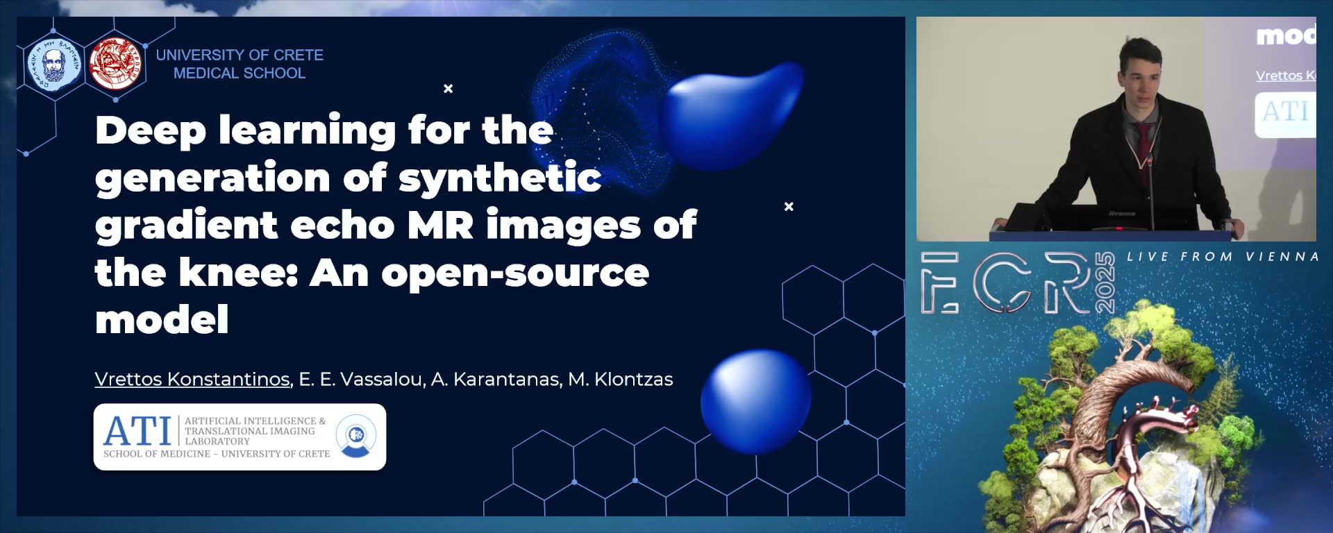
Deep learning for the generation of synthetic gradient echo MR images of the knee: An open-source model
08:00Konstantinos Vrettos, Heraklion / GR
2
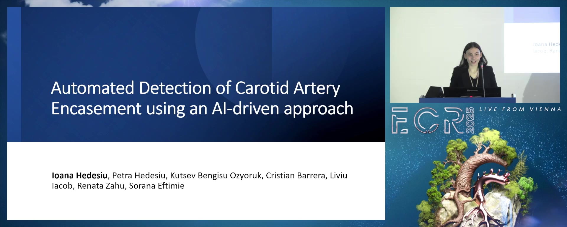
Automated Detection of Carotid Artery Encasement using an Artificial Intelligence Driven Approach
08:00Ioana Hedesiu, Liverpool / UK
3
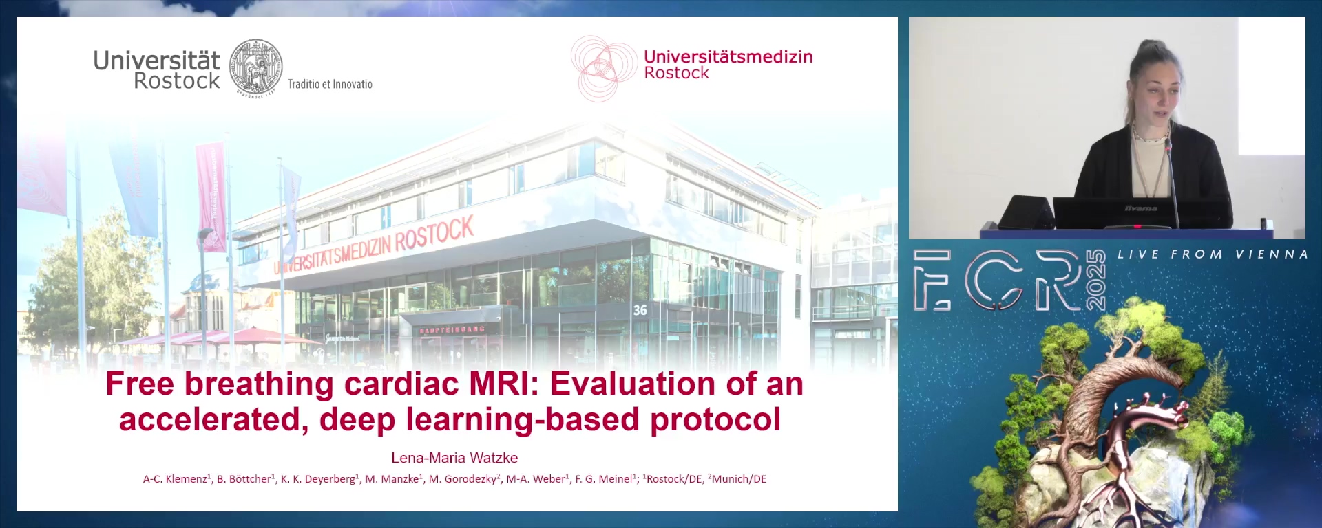
Free breathing cardiac MRI: Evaluation of an accelerated, deep learning-based protocol
08:00Lena-Maria Watzke, Rostock / DE
4
Detection of Pneumonia in Children through Chest Radiographs using Artificial Intelligence in a Low-Resource Setting: A Pilot Study
08:00Taofeeq Oluwatosin Togunwa, Ibadan / NG
5
Segmenting hippocampal subfields using deep learning reconstructed MRI images
08:00Nikolaus Maximilian Clodi, Linz / AT
6
Impact of bridging thrombolysis on outcome of mechanical thrombectomy and incidence of periprocedural emboli in ischemic stroke
08:00Dafne Vecchiet, Graz / AT
7
Evaluating Coronary Artery Calcium Burden and Prevalence across the Pooled Cohort Equations and American Heart Association PREVENT 10-Year Risk Models
08:00Gabrielle Sara Gershon, Atlanta / US
8
Enhancing diagnostic accuracy of subsegmental pulmonary embolism using CT pulmonary artery iodine subtraction imaging: a multi-reader, multi-case study
08:00Na Tan, Kunming / CN
9
Lateral Circumflex Femoral Artery embolization for the treatment of chronic hip pain related to osteoarthritis
08:00Maria Vittoria Paolucci, Rome / IT
10
Magnetic Resonance Imaging Based Radiomics for Predicting Pathogenetic Features and Survival in Rectal Cancer
08:00Bertalan Tóth, Budapest / HU
8 min
Deep learning for the generation of synthetic gradient echo MR images of the knee: An open-source model
Konstantinos Vrettos, Heraklion / Greece
Author Block: K. Vrettos, E. E. Vassalou, A. Karantanas, M. Klontzas; HERAKLION/GR
Purpose: A typical MR knee examination does not include T2*-w gradient echo (T2*W) images, but this sequence can be very useful in several cases, like pigmented villonodular synovitis (PVNS). This work proposes an open-source deep learning model that generates T2*W knee images from fat suppressed intermediate-weighted (FS IW) images.
Methods or Background: The model, which is based on the cycle-GAN architecture, was trained with 12,118 sagittal knee MR images and tested on an independent set of 2,996 images. Two experts evaluated the diagnostic interchangeability of the generated images for several findings including synovial blooming, chondrocalcinosis and chondral osteophytes. The image quality was further tested using objective metrics (PSNR, SSIM, NRMSE). The presence of artifacts was also analysed, together with their impact on the diagnostic value. A downloadable app, the code and model are available on GitHub.
Results or Findings: The synthetic images were deemed interchangeable with the originals, for all individual findings, with an ICC>0.95. The median PSNR, NRMSE and SSIM were 17.4, 0.5 and 0.5 respectively. There were four classes of artifacts: wrap-around-like (26/163 cases), object insertion/omission (11/163 cases), incomplete fat-suppression (120/163 cases) and geometrical distortion (86/163 cases), with a median 0 impact (no impact) on the diagnosis. Image synthesis for a single knee lasted <30sec.
Conclusion: An open-source cycle-GAN model has been trained to generate T2*W synthetic images of the knee. These images have demonstrated high image quality and high diagnostic value. Additionally, synthesizing the images took considerably less time than traditional acquisition, which can benefit both the patient and the clinician. While some artifacts have been observed, they have little to no impact on the diagnostic value of the images.
Limitations: Image quality metrics values could be improved, external validation is necessary and artifacts may appear.
Funding for this study: No funding was received for this study.
Has your study been approved by an ethics committee? Yes
Ethics committee - additional information: The study was approved by the Institutional Review Board.
8 min
Automated Detection of Carotid Artery Encasement using an Artificial Intelligence Driven Approach
Ioana Hedesiu, Liverpool / United Kingdom
Author Block: I. Hedesiu1, P. Hedesiu2, C. Barrera3, K. B. Ozyoruk4, L. Iacob2, R. Zahu2, S. Eftimie2; 1Liverpool/UK, 2Cluj-Napoca/RO, 3Atlanta, GA/US, 4Bethesda, MD/US
Purpose: Carotid artery encasement (CAE) can complicate surgical resection of head and neck squamous cell carcinomas (HNSCC), increasing the risk of surgery. Incomplete tumour excision due to the proximity of the carotid artery may impact the long-term outcomes by promoting tumour recurrence. This study aims to employ Artificial Intelligence for CAE detection on Computed Tomography (CT) images.
Methods or Background: The CT images and tumour masks were obtained from the National Cancer Institute CPTAC- HNSCC dataset. A hundred patients were included, totalling 1,367 tumour-present CT images. For the HNSCC segmentation, the pre-trained ResNet-50 backbone of a U-Net model was fine-tuned on the CT images from the dataset. The carotid segmentation utilised the MedSAM-2D foundation model, with a professional radiologist reviewing the selected bounding area. Both models returned the segmentation for the tumour and carotid, respectively. CAE was assessed by calculating the minimum distance and encasement angle between the masks returned by the models.
Results or Findings: During testing, the HNSCC segmentation model reached a mean Intersection over Union (IoU) score of 86%, with the tumour class achieving 73% accuracy and the non-tumour class 99%. Training required 54 epochs with early stopping. The carotid segmentation was best when employing the bounding-box method, achieving 79% accuracy. The two models succeeded in being applied to the same image to assess CAE.
Conclusion: An automated and accurate evaluation of CAE on CT images has been developed, demonstrating high accuracy and promising potential for future clinical applications.
Limitations: The dataset contains a limited number of CAE cases due to the rarity of this condition which may reduce the model’s accuracy.
Funding for this study: No funding
Has your study been approved by an ethics committee? Not applicable
Ethics committee - additional information: The study employed a publicly available dataset, qualifying for exemption from human research ethics review. The dataset use followed the terms of the TCIA-restricted license agreement.
8 min
Free breathing cardiac MRI: Evaluation of an accelerated, deep learning-based protocol
Lena-Maria Watzke, Rostock / Germany
Author Block: L-M. Watzke1, A-C. Klemenz1, B. Böttcher1, K. K. Deyerberg1, M. Manzke1, M. Gorodezky2, M-A. Weber1, F. G. Meinel1; 1Rostock/DE, 2Munich/DE
Purpose: Cardiac magnetic resonance imaging (cMRI) remains challenging for patients with shortness of breath and arrhythmias. In this study accelerated DL based real-time sequence in free-breathing (FB) and with breath-hold (BH) are compared in terms of volumetric results and acquisition time.
Methods or Background: In a prospective cohort study 25 healthy volunteers underwent a non-contrast cMRI examination at 1.5 T (SIGNA Artist, GE HealthCare, Milwaukee, U.S.A.). Short axis stack and long axis (2-, 3- and 4-chamber view) cine sequences of the left ventricle were acquired using Sonic DL™ (GE HealthCare) single heartbeat acquisition (FOV 34x34cm, matrix size 180x160, slice thickness 8mm) first in a BH then in FB. For a subset of 15 volunteers, the overall time of the FB and BH acquisitions was manually stopped with a stopwatch. Volumetric parameters were calculated using established post-processing software (cvi42, Circle Cardiovascular Imaging, Calgary, Canada). Wilcoxon signed rank sum test with a significance level of p<0.05 was performed for statistical analysis.
Results or Findings: Median acquisition time for BH and FB sequence stack was 178 sec [range: 138-270 sec] and 95 sec [64-140 sec], respectively (p<0.001). There was no statistically significant difference in end-diastolic (162.1 vs. 164.7 mL, p=0.35), end-systolic (65.6 vs. 62.6 mL, p=0.06) and stroke volume (95 vs. 98.7 mL, p=0.45) in FB compared to BH. There was a statistically significant but clinically irrelevant difference in ejection fraction (60.7 vs. 61.8 %, p=0.03).
Conclusion: The accelerated free-breathing protocol enables a 47% faster acquisition without compromising volumetric accuracy. Compared to conventional breath-hold techniques, it offers a more patient-friendly alternative, improving accessibility of cMRI for a wider range of patients.
Limitations: This study was conducted only on healthy individuals at a single MRI scanner from one vendor.
Funding for this study: None
Has your study been approved by an ethics committee? Yes
Ethics committee - additional information: The study was designed as a prospective, single-center cohort study and approved by the responsible institutional review board (Rostock)
8 min
Detection of Pneumonia in Children through Chest Radiographs using Artificial Intelligence in a Low-Resource Setting: A Pilot Study
Taofeeq Oluwatosin Togunwa, Ibadan / Nigeria
Author Block: T. O. Togunwa, A. Babatunde, R. B. Olatunji, O. E. Fatade, G. I. Ogbole, A. Falade; Ibadan/NG
Purpose: Pneumonia remains a leading cause of death in under-5 children in low- and middle-income countries (LMICs), exacerbated by limited diagnostic imaging expertise. Artificial intelligence (AI) shows potential in improving pneumonia diagnosis from chest radiographs (CXRs). However, most models lack external validation on prospective clinical data from LMICs. This study aims to develop and validate an AI model for childhood pneumonia detection in Nigeria.
Methods or Background: In a multi-center cross-sectional study in Ibadan, Nigeria, CXRs were prospectively collected from two radiology departments via cluster sampling (November 2023–August 2024). A VGG-19-based AI model was trained and validated (internal test) using open-source paediatric CXR dataset from the USA; to classify local CXRs as either normal or pneumonia. Two blinded radiologists provided consensus classification as the reference standard. The model's accuracy, precision, recall, F1 score, and AUC were evaluated.
Results or Findings: The AI model was developed on 5,232 open-source paediatric CXRs, divided into training (1,349 normal, 3,883 pneumonia) and internal test (234 normal, 390 pneumonia) sets, and externally tested on 190 radiologist-classified CXRs (93 normal, 97 pneumonia). The model achieved 86% accuracy, 0.83 precision, 0.98 recall, 0.79 F1 score, and 0.93 AUC on the internal test, and 58% accuracy, 0.62 precision, 0.48 recall, 0.68 F1 score, and 0.65 AUC on the external test (95% CI).
Conclusion: This study demonstrates AI’s potential in diagnosing childhood pneumonia with reasonable performance on internal tests. However, external test results highlight the need for further AI development to enhance AI generalizability across diverse clinical settings.
Limitations: The model's lower performance on external tests highlights challenges in applying AI across diverse healthcare settings, likely due to differences in imaging protocols and equipment. Prioritizing robust, region-specific African datasets is key to sustainable AI development in the region.
Funding for this study: Funding was provided by Center for Policy Impact in Global Health, Duke Global Health Institute
Has your study been approved by an ethics committee? Yes
Ethics committee - additional information: The study was approved by the University of Ibadan/ University College Hospital ethics committee (UI/EC/23/0651)
8 min
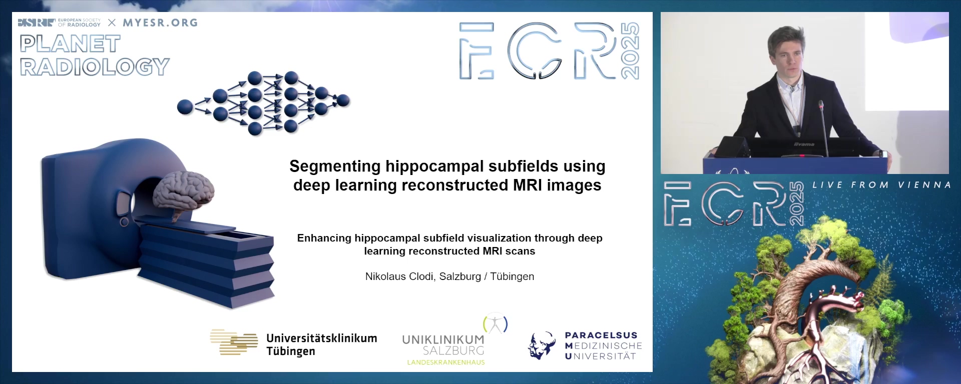
Segmenting hippocampal subfields using deep learning reconstructed MRI images
Nikolaus Maximilian Clodi, Linz / Austria
Author Block: N. M. Clodi1, B. Bender2, G. Hecke1, G. Gohla2, T-K. Hauser2, P. Ghibes2, K. D. Hergan1, U. Ernemann2, A. Estler2; 1Salzburg/AT, 2Tübingen/DE
Purpose: Assessing hippocampal pathology in epilpesia can be challenging. To enhance diagnostic accuracy, deep learning image reconstruction, standardized imaging protocols, and image post-processing methods are proposed. This study compares T2 TSE DRB Sequences and standardized T2 TSE Sequences in hippocampal segmentation and volumetry using FreeSurfer.
Methods or Background: FreeSurfer (Version 7.4.1) is utilized for segmenting hippocampal subregions between the T2 TSE DRB and T2 TSE sequences in 36 subjects (mean age 39±14 years; 21 men, 15 women). The segmented volumes are then compared using a two-tailed t-test. Furthermore, a 95% confidence interval, derived from the mean percentage difference in hippocampal volumes among healthy individuals, is provided to assist in the objective assessment of hippocampal pathology.
Results or Findings: When comparing all hippocampal segments combined between the T2 TSE DRB and T2 TSE sequences, the volumes were identical. However, individual segment analysis showed significant volume differences in the CA1-Body (p=0.003; cohen's d=0.0827), CA4-Body (p=0.002; d=0.1501), and whole_hippocampal_body (p=0.012; d=0.0149) region of the right hippocampus. No other individual region was otherwise different. Additionally, pathological volume changes between the left and right hippocampus in a single subject could be identified using the proposed 95% confidence interval, tested using diseased.
Conclusion: The small number of significant differences and the low effect size demonstrate that novel deep learning-based sequences are not inferior to conventional sequences. Novel T2 TSE DRB sequences can therefore be used for segmentation of the hippocampus without uncertainty. Additionally, the proposed 95% interval could contribute to a more objective description of hippocampal pathology.
Limitations: Small sample size
Retrospective study
Single center study
Publication bias
Funding for this study: Supported by Fortüne-Program of the medical faculty of Tübingen and by the Prof. Dr. Peter und Jytte Wolf – Stiftung für Epilepsie
Has your study been approved by an ethics committee? Yes
Ethics committee - additional information: This retrospective monocentric study received approval from the local review board (University Tübingen), and informed consent was waived under the code 161/2023B01. The study adhered to the principles outlined in the Declaration of Helsinki guidelines and recommendations.
8 min
Impact of bridging thrombolysis on outcome of mechanical thrombectomy and incidence of periprocedural emboli in ischemic stroke
Dafne Vecchiet, Graz / Austria
Author Block: D. Vecchiet, C. Reiter, M. Kneihsl, H. Deutschmann; Graz/AT
Purpose: Mechanical thrombectomy is the standard of care for acute treatment of ischemic stroke due to large vessel occlusion. Recent studies suggest improved functional outcome by combining mechanical thrombectomy with intravenous bridging thrombolysis. Thrombolysis may, however, decrease thrombus stability, potentially increasing periprocedural embolisms. The purpose of this study was to assess the impact of bridging thrombolysis on mechanical thrombectomy outcome and periprocedural emboli incidence in ischemic stroke.
Methods or Background: Patients who underwent mechanical thrombectomy with or without bridging thrombolysis for treatment of ischemic stroke at our institution between 2016 and 2021. were retrospectively analyzed. Angiographic modified treatment in cerebral infarction (mTICI) score, modified Rankin Scale after 90 days (mRS90), frequency of periprocedural embolisms, and the impact of technique (aspiration vs. stent retriever) were evaluated. Successful angiographic outcome was defined as mTICI≥2b, and good functional outcome as mRS90≤2.
Results or Findings: A total of 450 patients were included. Combination of mechanical thrombectomy and bridging thrombolysis resulted in significantly higher rates of successful angiographic outcome (mTICI≥2b 93% vs. 86%, p=0.017) and good functional outcome (mRS90≤2 34% vs. 45%, p=0.018) compared to thrombectomy alone. No difference in occurrence of periprocedural embolisms was observed between groups (29% vs. 29%, p=0.906). However, in the aspiration subgroup, bridging thrombolysis increased periprocedural embolisms (17% vs. 53%, p=0.039 ), though not significantly affecting functional outcome (mRS90≤2: 33% vs. 26%, p=0.126).
Conclusion: Bridging thrombolysis enhances the outcome of mechanical thrombectomy for treatment of ischemic stroke. The observed higher incidence of periprocedural embolism in patients treated with aspiration and bridging thrombolysis might be explained by limited availability of balloon guide catheters during the investigated timeframe.
Limitations: Retrospective study, small subgroup size.
Funding for this study: None
Has your study been approved by an ethics committee? Yes
Ethics committee - additional information: Obtained
8 min
Evaluating Coronary Artery Calcium Burden and Prevalence across the Pooled Cohort Equations and American Heart Association PREVENT 10-Year Risk Models
Gabrielle Sara Gershon, Atlanta / United States
Author Block: G. S. Gershon, Y. Yang, A. Razavi, L. Sperling, C. Onnis, C. N. De Cecco, M. van Assen; Atlanta, GA/US
Purpose: The purpose of this study is to compare coronary artery calcium (CAC) burden across risk groups defined by the Pooled Cohort Equation (PCE) and the PREVENT risk calculator, to assess how CAC improves risk stratification for atherosclerotic cardiovascular disease (ASCVD) and heart failure (HF) beyond these models.
Methods or Background: The 2013 PCE has served to assess ASCVD risk. In 2023, the PREVENT risk calculator was developed considering the cardiovascular-kidney-metabolic health paradigm. CAC, measured on non-contrast cardiac computed tomography (CT), improves ASCVD risk stratification. The prevalence of CAC burden across PCE versus PREVENT risk groups is unknown. This retrospective study included 7,610 asymptomatic individuals with CAC CT from 2010-2023 at Emory. The 10-year clinical risk by PCE and base PREVENT were retrospectively calculated. CAC was quantified using the Agatston method. CAC scores across risk groups were compared with Kendall Tau tests, using CAC (0, 1-99, 100-299, ≥300) and PCE/PREVENT (low<5, borderline-intermediate 5-19, high≥20%).
Results or Findings: The mean age was 57, 41% were women, and 29% were non-White. PCE classified 52.5% (n=3,997) low, 41% (n=3,118) borderline-intermediate, and 6.5% (n=495) high-risk, whereas ASCVD PREVENT classified 72.6% (n=5,523) low, 27.1% (n=2,059) borderline-intermediate, and 3.7% (n=28) high-risk. CAC distribution was heterogeneous across risk categories within both PCE and all PREVENT, and between PCE and all PREVENT (p<0.001). CAC prevalence was 71% and 79% for individuals with borderline-intermediate risk according to PCE and ASCVD PREVENT, respectively, and 72% for CVD and 80% HF PREVENT.
Conclusion: The prevalence of CAC was similar across calculated PCE and PREVENT risk groups, including individuals with borderline-intermediate risk. These results underline the continued utility of CAC in improving risk stratification beyond PCE and PREVENT.
Limitations: No limitations were identified.
Funding for this study: Funding was provided by American Heart Association-- Student Scholarship in Cardiovascular Disease.
Has your study been approved by an ethics committee? Yes
Ethics committee - additional information: IRB at Emory University
8 min
Enhancing diagnostic accuracy of subsegmental pulmonary embolism using CT pulmonary artery iodine subtraction imaging: a multi-reader, multi-case study
Na Tan, Kunming / China
Author Block: N. Tan; Kunming/CN
Purpose: CT pulmonary artery iodine subtraction imaging (LSIM) visualizes perfusion abnormalities associated with embolism-induced bloodflow reduction. Our aim is to enhance SSPE diagnostic accuracy by integrating CTPA with LSIM and expanding its applicability beyond radiologiststo clinical physicians.
Methods or Background: In this prospective study conducted from July 2023 to July 2024, 1,211 patients with suspected PE underwent CTPA using a Canon 320 detector CT or Siemens Definishion Flash. Motion-corrected iodine subtraction images (enhanced CT minus pre-enhanced CT) were generated. Ground truth for SSPE was established by expert consensus in a total of 132 cases. The observers included 2 clinicians each from emergency medicine, respiratory medicine and vascular surgery, and 6 radiologists of varying experience. They assessed 260 randomly selected patients, equally divided between SSPE positive and negative cases (1:1 ratio), using stand-alone CTPA and iodine subtraction CT for PE diagnosis.
Results or Findings: Combining LSIM iodine subtraction imaging significantly enhances SSPE detection rate (CTPA [0.74], LSIM [0.96], p = .025); LSIMmarkedly improves diagnostic accuracy for junior radiologists and clinical physicians (κ=0.95, specificity 0.93), compared to CTPA (p = .015);average reading time with LSIM decreases (CTPA [153 s] vs LSIM [110 s], p = .06); average AUC of observers shows substantial improvement(iodine subtraction CT [0.94 (0.90, 0.99)] vs CTPA [0.88 (0.83, 0.92)], P = .02).
Conclusion: LSIM enhances the detection rate and diagnostic accuracy of subsegmental pulmonary embolism (SSPE), particularly notableamong clinical physicians, while also reducing time to diagnosis.
Limitations: This study’s primary limitation lies in its short duration and single-center design, which may affect the generalizability of the results across diverse clinical settings and populations. Further multicenter trials are recommended to validate the findings.
Funding for this study: No
Has your study been approved by an ethics committee? Not applicable
Ethics committee - additional information: Ethics Committee of the Yan'an Hospital of Kunming Medical University
8 min
Lateral Circumflex Femoral Artery embolization for the treatment of chronic hip pain related to osteoarthritis
Maria Vittoria Paolucci, Rome / Italy
Author Block: M. V. Paolucci, M. Rossi, M. A. Tipaldi, E. Ronconi, G. Orgera, A. Zolovkins; Rome/IT
Purpose: To evaluate the safety and effectiveness of the lateral circumflex femoral artery embolization employing degradable starch microspheres to treat chronic pain due to osteoarthritis.
Methods or Background: A cohort of seven patients, with a median age of 64 years ( range 45-84 years) has been enrolled in a single-center, single arm, prospective study. Outcomes were assessed adopting the Western Ontario and McMaster Universities Osteoarthritis Index (WOMAC) and Visual Analogue Scale (VAS) at baseline, 48 hours, 1 month, 3 months and 6 months post-procedure. Major inclusion criteria were: pain symptoms unresponsive to conservative treatments, osteoarthritis according to Kellgren-Lawrence grading system (grade 1 to 3), clear signs of osteoarthritis on MRI, VAS >5. The embolization was carried out using degradable starch microspheres EmboCept S 50 µm.
Results or Findings: LCFA embolization resulted in significant pain reduction right after the procedure with VAS scores improving from 8.0 ± 1.2 pre-procedure to 2.5 ± 1.3 at 6 months post-procedure. WOMAC scores demonstrated improved joint function and quality of life, with marked reduction in stiffness and physical dysfunction. There were no significant differences in the reported improvements from patients at 48 hours, 1 month, 3 months and 6 months post-procedure, indicating that the therapeutic benefits are sustained over time. No major complications were reported following the procedure.
Conclusion: LCFA embolization appears to be a promising treatment for chronic hip pain related to osteoarthritis. This procedure offers a safe and effective option, particularly for those patients who seek to avoid surgery. However, additional long-term research is required to confirm its lasting effects and optimize criteria for selecting suitable patients.
Limitations: Limited sample and short follow-up period.
Funding for this study: No funding for this study
Has your study been approved by an ethics committee? Yes
Ethics committee - additional information: Approved by an ethics committee
8 min
Magnetic Resonance Imaging Based Radiomics for Predicting Pathogenetic Features and Survival in Rectal Cancer
Bertalan Tóth, Budapest / Hungary
Author Block: B. Tóth, A. Tarnoki, D. L. Tarnoki; Budapest/HU
Purpose: This study aims to evaluate the potential of Magnetic Resonance Imaging based radiomics in predicting key pathological features and long-term survival outcomes in rectal cancer patients.
Methods or Background: A retrospective study was conducted on 510 rectal cancer patients treated between 2015 and 2019. The inclusion criteria required pre-therapeutic MRI performed on a Discovery MR750W 3.0T machine and known KRAS mutation status. Forty-seven patients met the criteria. MRI sequences included T1-weighted, T2-weighted fat-saturated , high-resolution T2-weighted, and diffusion-weighted imaging. Manual segmentation was used with the help of 3DSlicer. Radiomic features were extracted using PyRadiomics, and machine learning models were developed using XGBoost and LightGBM classifiers. Feature selection was performed using Sequential Feature Selector and Minimum Redundancy Feature Selection.
Results or Findings: The model for KRAS mutation status achieved an AUC of 0.7475 (training) and 0,75 (testing). Lymph node invasion prediction had an AUC of 0.7892 (training) and 0.7984 (testing). Vascular invasion prediction yielded an AUC of 0.6989 (training) and 0.7143 (testing). The 5-year survival prediction model showed an AUC of 0.7848 (training) and 0.7750 (testing). Metastasis prediction achieved an AUC of 0.6627 (training) and 0.6857 (testing).
Conclusion: MRI-based radiomics demonstrates significant potential in predicting key pathological features and long-term survival outcomes in rectal cancer patients. Integrating multimodal imaging data and clinical information, along with automated segmentation techniques, could further enhance model accuracy and clinical utility.
Limitations: The limitations of the study are the reliance on single-modality imaging, without incorporating additional modalities like CT or PET/CT, which limits the predictive power of our models. Furthermore, the relatively small sample size and the manual segmentation process create challenges for generalizability and efficiency.
Funding for this study: Funding was provided by the National Tumor Biology Laboratory project (NTL-19) and the National Artificial Intelligence Laboratory (RRF-2.3.1-21-2022-00004). The project was implemented with the support of the Ministry of Culture and Innovation from the National Research, Development and Innovation Fund, based on a funding agreement with the National Research, Development and Innovation Office (National Laboratories Program – National Tumor Biology Laboratory – 2022-2.1.1-NL-2022-00010).
Has your study been approved by an ethics committee? Yes
Ethics committee - additional information: The research ("Radiomics Analysis of Breast Tumors, Colorectal, Head and Neck, and Ovarian Cancers") was ethically approved by the Regional Research Ethics Committee and the Scientific and Research Ethics Committee of the Health Science Council (07464-3/2023/EÜIG).