Research Presentation Session: Radiographers
RPS 2214 - A focus on MRI and mammography practice and care
RPS 2214-3
7 min
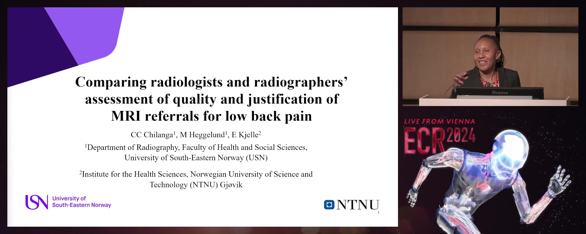
Comparing radiologists' and radiographers' assessment of quality and justification of lower back MRI referrals
Catherine Chilute Chilanga, Drammen / Norway
- Author Block: C. C. Chilanga1, M. Heggelund1, E. Kjelle2; 1Drammen/NO, 2Gjøvik/NO
Purpose: This study aimed to compare radiologists' and radiographers' assessment of the quality of Magnetic Resonance Imaging (MRI) lumbosacral spine referrals for low back pain (LBP).
Methods or Background: A total of N=300 lumbosacral spine MRI referrals for LBP for adults aged 16–90 years were obtained. A registration form was designed consisting of seven statements on relevant information in the referral rated on a three-point scale: 'Yes', 'No' or 'Uncertain'. In addition, the referral was rated as 'Justified', 'Unjustified' or 'Need more information'. The registration form was pilot-tested on two radiologists to check for validity and reliability. Four radiologists and two radiographers were recruited and assigned the task to assess the same 300 referrals individually and complete the registration form. STATA Statistics packages were utilised to perform mixed model analysis to show variability in ratings between groups.
Results or Findings: Overall, an average of 65% (n=196) of the referrals were assessed as justified. There was a difference between radiographers and radiologists in the assessment of referral justification with
- 5% (95% CI [69.4%-77.7%]) and 58.4% (95% CI [54.5%-62.1%]) rated 'Justified' respectively. This variation was statistically significant (p-value <0.001). In referral quality, an average of 7% (n=22) received a ‘good’ quality score. Compared to the radiologists, the radiographers rated a statistically significant (p<0.001) higher percentage of the referrals as good quality, with radiologists rating 6.2% (95% CI [4.5%-8.0%] as good quality and radiographers rating 10% (95% CI [7.3%-12.6%]) to be of good quality. Conclusion: The radiographers rated more LBP MRI referrals as 'Justified' and gave the referrals an overall higher quality score than radiologists. Limitations: The limitations of the study are that data is only from a few raters, with an added unequal amount representation of professional groups, limiting generalising of the findings. Funding for this study: Funding was provided by The Research Council of Norway (Project number 302503). Has your study been approved by an ethics committee? Yes Ethics committee - additional information: The study was approved by the Regional Committees for Medical and Health Research Ethics (REK) reference number 378396 and the Norwegian Agency for Shared Services in Education and Research (SIKT) reference
RPS 2214-4
7 min
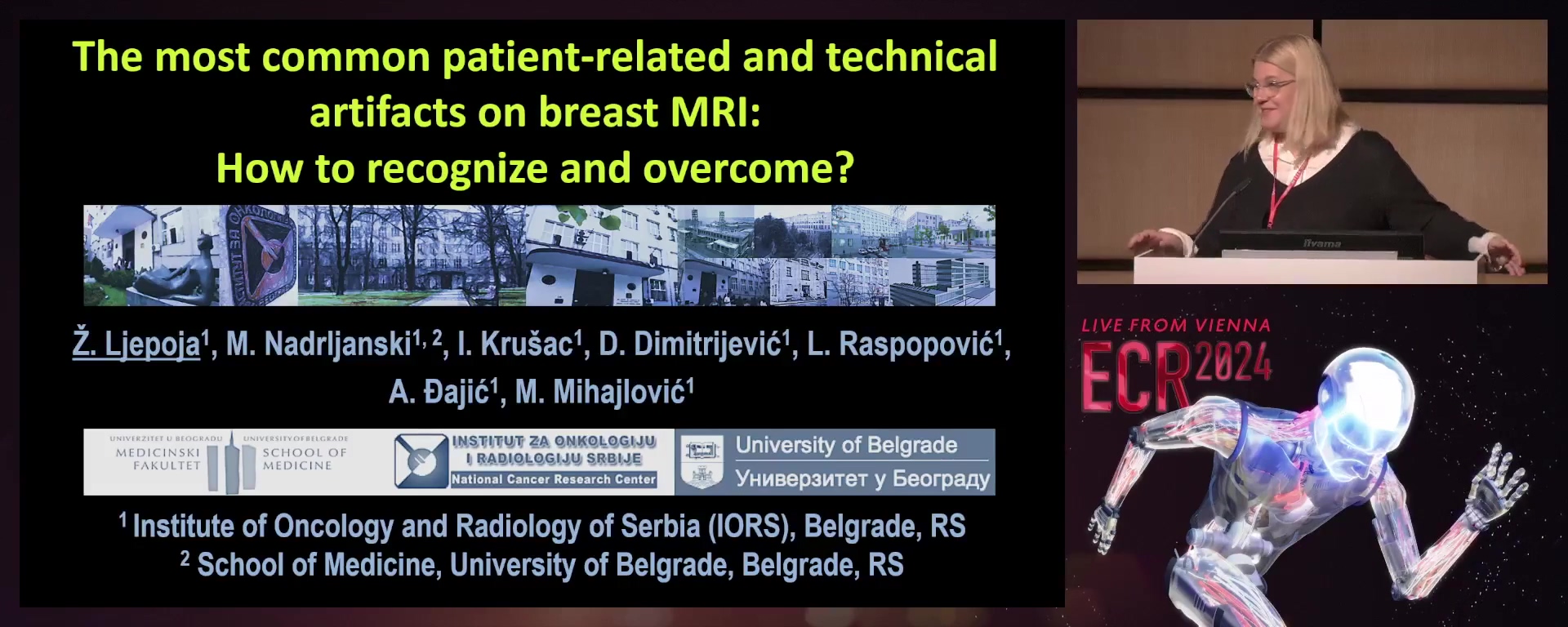
The most common patient-related and technical artefacts on breast MRI: how to recognise and overcome?
Zeljka Ljepoja, Belgrade / Serbia
- Author Block: Z. Ljepoja, M. M. Nadrljanski, I. B. Krušac, D. Dimitrijevic, L. Raspopovic, A. Djajic, M. Mihajlovic; Belgrade/RS
Purpose: This study aimed to recognise the most common artefacts and misinterpretations of the objects in the field of view: patient-related (motion artefacts, artefacts due to positioning, metal artefacts) and technical (zipper artefacts, wrap-around artefacts, chemical shift artefacts and zebra artefacts).
Methods or Background: 300 consecutive breast MRI exams, all realised with a full diagnostic protocol (T2W-STIR, T2W-TSE, T1W-TSE, FLASH 3D with the application of the same contrast medium: gadobutrol) on
- 5T MRI unit (realised: October 2020 –October 2023) were analysed for the preselected sets of artefacts (patient-related and technical). Results or Findings: Artefacts were detected on
- 67% of all analysed MRI breast exams. Artefacts predominantly belonging to patients were 84.74%. Motion artefacts were 45.76%; artefacts due to positioning were 23.73%; metal artefacts were 15.25%. Only 15.25% of all artefacts were technical artefacts. Zebra artefacts were 5.08%; wrap-around artefacts were 3.39%; zipper artefacts were 3.39%, and chemical shift artefacts were 3.39%. Significantly more patient-related artefacts were detected (motion artefacts, p=4.45e-7), with the pval for distribution of 0.046, favouring the presence of motion artefacts. The diagnostic interpretation was affected by patient-related motion artefacts with 7 exams being rescheduled and repeated, i.e. 11.86% of all detected artefacts. Conclusion: Technologists and radiologists need to recognise and understand the artefacts on breast MRI in order to provide the satisfactory and permanent quality of the images. Adequate patient preparation is important for the adequate image quality. Limitations: This was a retrospective analysis. Funding for this study: No funding was received for this study. Has your study been approved by an ethics committee? Not applicable Ethics committee - additional information: This was technical research, exempted from the decision of the ethical committee.
RPS 2214-5
7 min
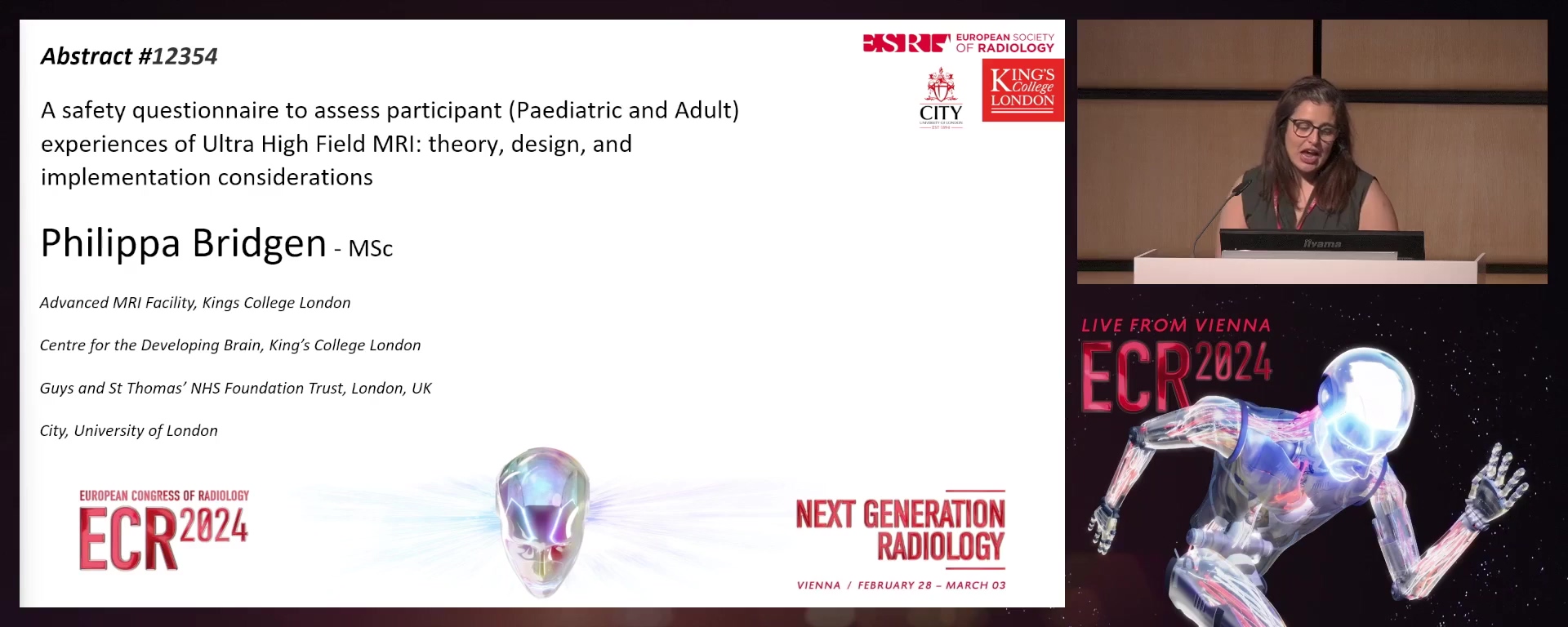
A safety questionnaire to assess participant (paediatric and adult) experience of ultra-high-field MRI: theory, design, and implementation considerations
Philippa Bridgen, London / United Kingdom
Author Block: P. Bridgen1, K. Colford1, B. Hansson2, I. M. Björkman-Burtscher3, T. Arichi1, S. Malik1, S. Giles1, C. Malamateniou1, G. Turner1; 1London/UK, 2Lund/SE, 3Gothenburg/SE
Purpose: The aim of this research was to create a robust, user-informed, evidence-based questionnaire for patient safety aspects at 7T, using local expertise and previous literature.
Methods or Background: 7T MRI increases signal-to-noise-ratio (SNR) and improves contrast in comparison to standard magnetic field strengths, giving the potential for additional information clinically. However, patients can experience transient sensory effects during 7T examinations, which may impact patient experience and acceptability. Previous research has primarily focused on adult perception, with children perceptions so far only being extrapolated from 3T data. Expanding these questionnaires to children to gain further necessary information is needed. Literature searches were carried out looking for both MRI transient effects and questionnaire designs at all MRI field strengths. Content analysis of 32 articles was completed to identify common themes, directed the subject of questions asked, following the patients’ MRI-scan journey. Answer formats included free comments and 5-point Likert scales. Questionnaires were adapted to be age-appropriate for 5-8 year and 8-11 olds, and adults. Language level was verified using the Flesch-Kincaid method. All questions were piloted (n=10) to gain feedback from intended users on content, design, and flow.
Results or Findings: Three comparable age-appropriate questionnaires were constructed, reflecting identified common themes from literature. Pictures aided understanding for children aged 5-8 years old. Questionnaires were divided into six sections: initial overview, positioning, entering, during the scan, exiting and post scan.
Conclusion: Newly designed questionnaires will allow a better understanding of how children or adults may experience 7T MRI and enhance safety strategies. Evidence collected from future use will support change of current practice.
Limitations: 7T is self-limiting due to the narrow scope, as such there is the potential for a lack of diversity of participants.
Funding for this study: No funding was obtained for this study.
Has your study been approved by an ethics committee? Yes
Ethics committee - additional information: Ethical approval granted from Committee of the School of Health Sciences, City, University of London under ETH2223-1703
RPS 2214-6
7 min
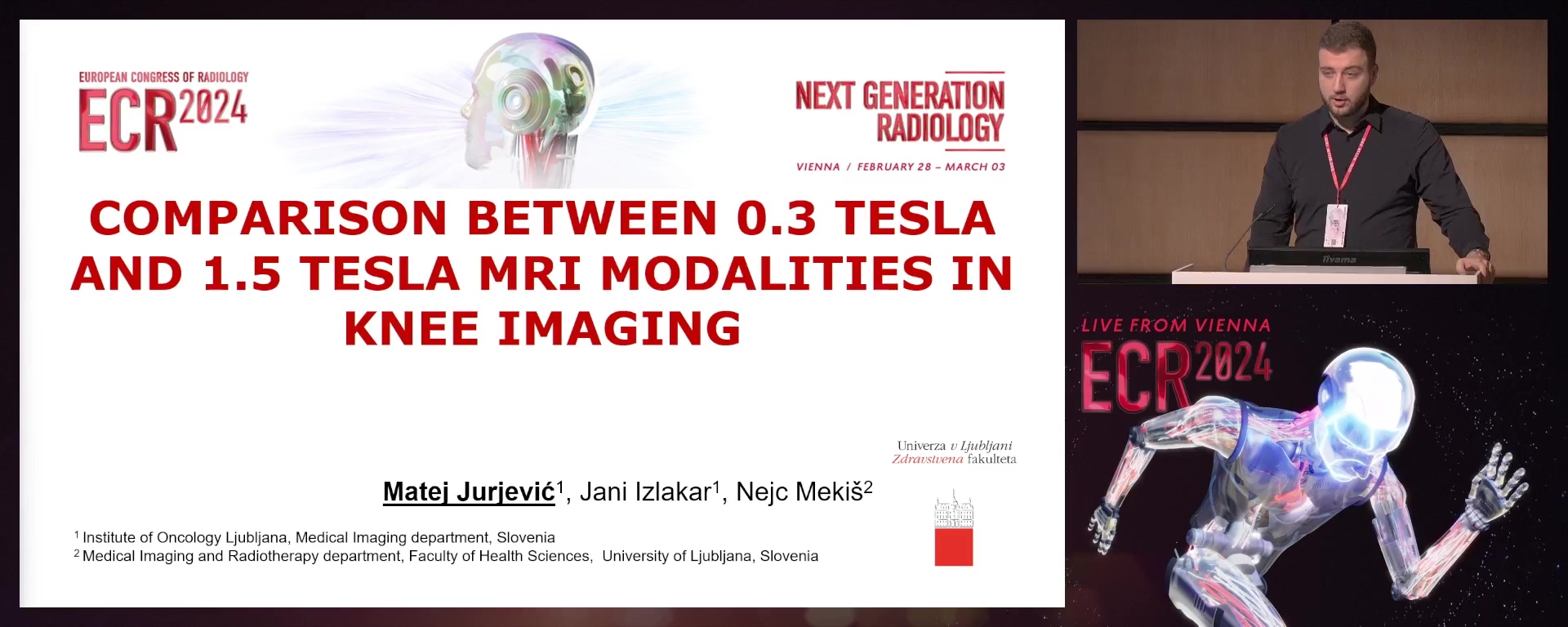
Comparison between 0.3 Tesla and 1.5 Tesla MRI modalities in knee imaging
Matej Jurjević, Kisovec / Slovenia
- Author Block: M. Jurjević, N. Mekis, J. Izlakar; Ljubljana/SI
Purpose: This study compared knee imaging at
- 3T and 1.5T field densities. We examined differences in signal-to-noise ratio (SNR), contrast-to-noise ratio (CNR), and image quality assessed by three experienced radiologists. Methods or Background: A sample of 25 left knees was examined using both MR devices with a 3 mm slice thickness. The volunteers were healthy and had no previous knee injuries. SNR and CNR measurements were performed on the medial meniscus, the distal part of the femur, articular cartilage, and the background. In the second part of the study, three radiologists assessed the image quality of the ACL, PCL, menisci, articular cartilage, and the overall image. Results or Findings: In the SNR and CNR measurements, we recorded statistically significant differences in the area of the medial meniscus in favor of the
- 5T modality for both SNR (p < 0.001) and CNR (p < 0.001). Similar results were observed in the area of the distal part of the femur, with better values for the 0.3T modality for both SNR (p < 0.001) and CNR (p < 0.001). However, in the measurements of the distal femur, we did not find statistically significant differences in the values of SNR (p = 0.677) and CNR (p = 0.861). For all selected structures, radiologists rated the 1.5T modality higher, and this is supported by the statistically significant differences. The agreement between radiologists was moderate for the ratings of menisci, articular cartilage, and the overall image, and poor for the quality of cruciate ligaments. Conclusion: Our results show superior image quality on
- 5T MRI, while indicating the potential for 0.3T open-type scanners in knee diagnostics. Limitations: Smaller FOV in one plain on
- 3T to reduce wrap-around artifact. Surface array coil used on 1.5T scanner. Funding for this study: Funds for this study were obtained from the Institute of Oncology Ljubljana, Medisken Trbovlje. Has your study been approved by an ethics committee? Yes Ethics committee - additional information: This study received ethical approval from the National Medical ethics committee, approval number: 0120-356/2022/
RPS 2214-7
7 min
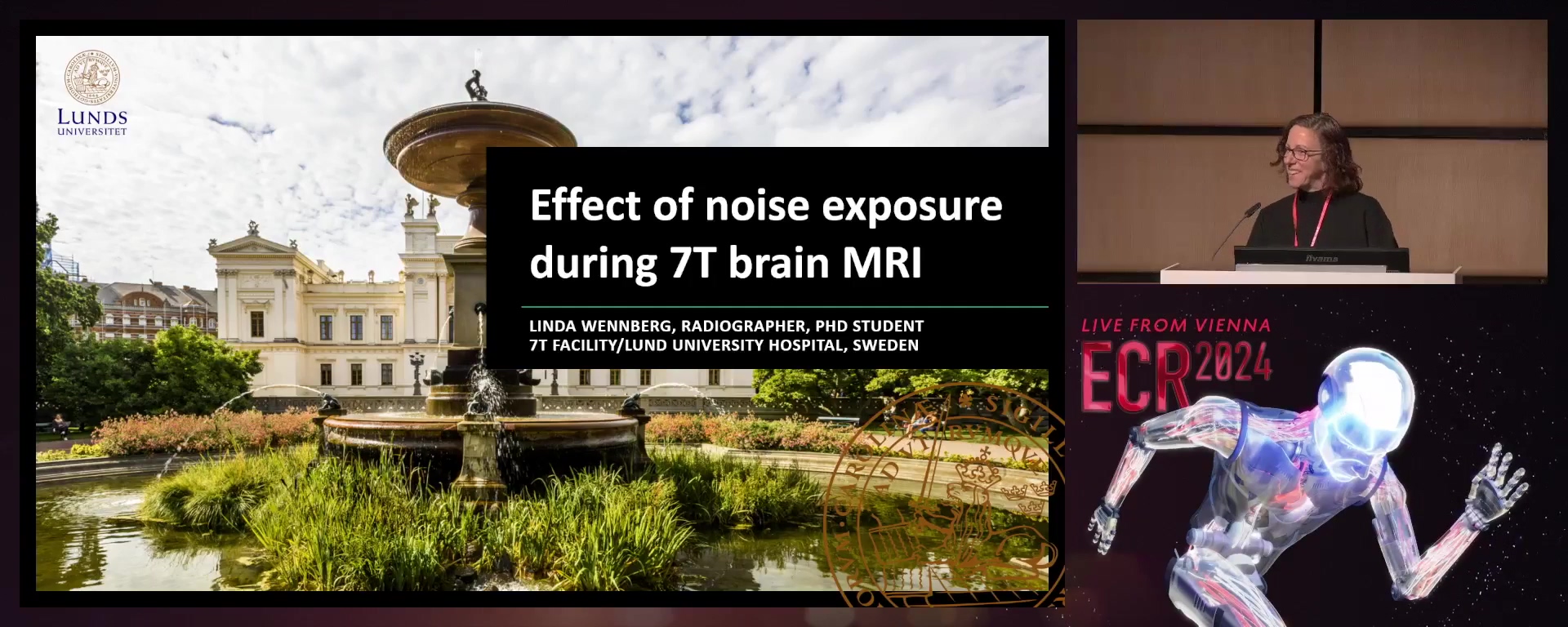
Effect of noise exposure during 7 Tesla MRI
Linda Maria Viviann Wennberg, Lund / Sweden
- Author Block: L. M. V. Wennberg, P. C. Maly Sundgren, S. Waechter, J. Brännström, A. Jönsson, B. Hansson, J. Mårtensson; Lund/SE
Purpose: This study aimed to assess the potential impact of noise exposure during a 7 Tesla (T) brain MRI in healthy adults.
Methods or Background: Excessive noise can harm the cochlear outer hair cells, leading to auditory damage. In this study, we used otoacoustic emission (OAE) to evaluate the effects of noise exposure in 39 healthy adults after a 7T MRI scanning session utilizing currently accepted hearing protection. The participants were enrolled in a research project involving two one-hour MRI scanning sessions on the same day. OAE assessments were performed before and after each scan, with a follow-up performed one week later.
Results or Findings: Our analysis revealed no significant differences in outer hair cell function between the baseline measurements and the first MRI scan. A significant difference was observed at
- 5 kHz and 2 kHz in the left ear, as well as at 4 kHz in the right ear after the second MRI scan. However, the follow-up OAE measurement showed no significant difference compared to the baseline at any of the frequencies in either ear. Conclusion: Our study's findings suggest no lasting effects on outer hair cell function in adults who undergo two one-hour MRI scanning sessions in a single day while using appropriate hearing protection. Limitations: It should be noted that the participants in this study were restricted to healthy young adults who met specific criteria. Therefore, further investigations involving a more diverse group of individuals would be beneficial to gain a more comprehensive understanding of the impact of 7T MRI scanning sessions on different populations. Funding for this study: This work was supported by grants from the Swedish Research Council (2017-00896) and the LMK Foundation. Both were awarded to Johan Mårtensson. Has your study been approved by an ethics committee? Yes Ethics committee - additional information: This study was approved by the local ethics committee (registration numbers 2019-05387, 2016/126, and 2020-06907).
RPS 2214-8
7 min
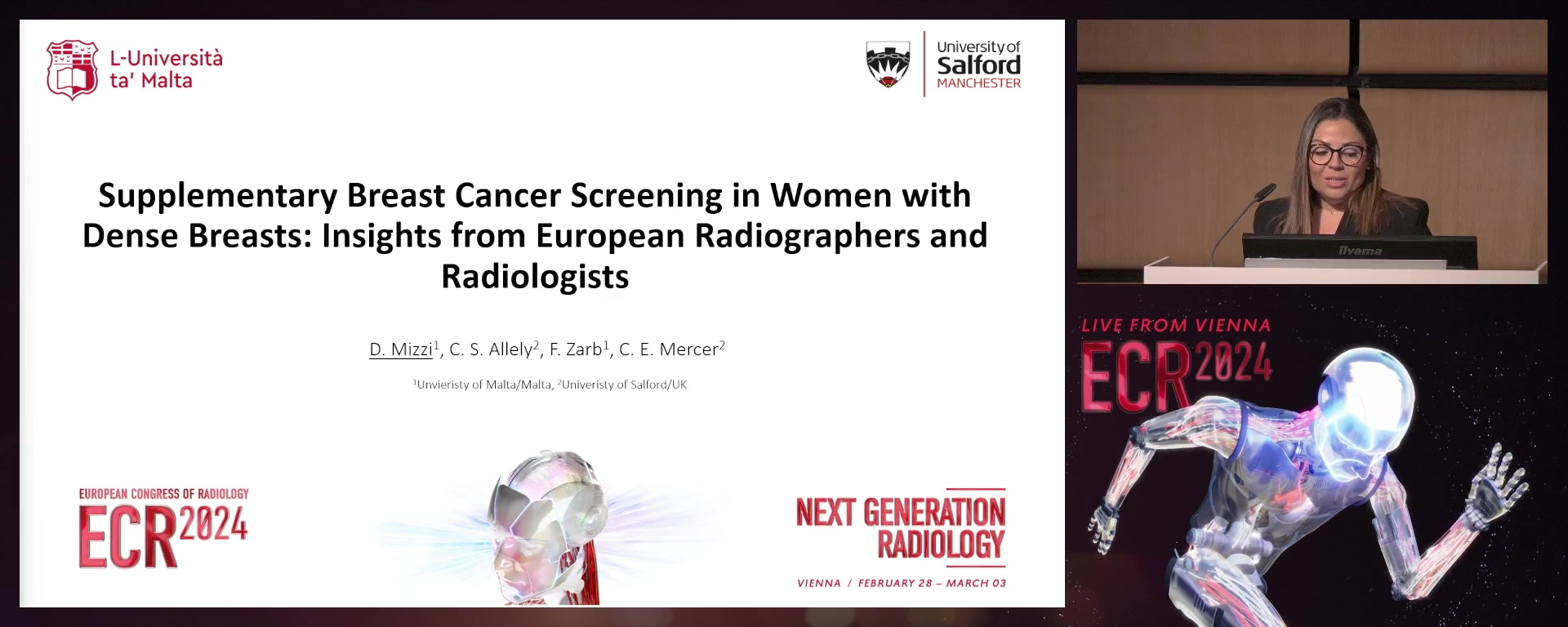
Supplementary breast cancer screening in women with dense breasts: insights from European radiographers and radiologists
Deborah Mizzi, Msida / Malta
Author Block: D. Mizzi1, C. Allely2, F. Zarb1, C. Mercer2; 1Msida/MT, 2Salford/UK
Purpose: This study explored the understanding of challenges and requirements for implementing supplementary breast cancer screening for women with dense breasts among clinical radiographers and radiologists in Europe.
Methods or Background: Fourteen semi-structured online interviews were conducted with European clinical radiologists (n=5) and radiographers (n=9) specializing in breast cancer screening from eight different countries including United Kingdom, Malta, Italy, the Netherlands, Greece, Finland, Denmark and Switzerland. The interview schedule comprised questions regarding professional background and demographics and 13 key questions divided into six subgroups, namely supplementary imaging; training; resources and guidelines; challenges; implementing supplementary screening and the women’s perspective of supplementary imaging. Data analysis followed the six phases of reflexive thematic analysis.
Results or Findings: Six significant themes emerged from the data analysis: understanding and experiences of supplementary imaging for women with dense breasts; challenges and requirements related to training among clinical radiographers and radiologists; awareness among radiographers and radiologists of guidelines on imaging women with dense breasts; challenges to implement supplementary screening; predictors of Implementing supplementary screening and Views of radiologists and radiographers on women's perception towards supplementary screening.
Conclusion: The interviews with radiographers and radiologists provided valuable insights into the challenges and potential strategies for implementing supplementary breast cancer screening. These challenges included cost and logistics problems and patient and staff related challenges. Implementing multifaceted solutions such as Artificial Intelligence integration, specialised training and resource investment can address these challenges and promote the successful implementation of supplementary screening in women with dense breasts. Further research and collaboration are needed to refine and implement these strategies effectively.
Limitations: The data collection period coincided with the reopening of screening units after COVID-19 closures. During this period, participants were exceptionally busy, which limited their availability to partake in the study.
Funding for this study: This work is part of a PhD programme which is part-financed by the Tertiary Education Scholarship Scheme (TESS), Government of Malta (TESS Contract MEDE 417/2018/61).
Has your study been approved by an ethics committee? Yes
Ethics committee - additional information: Ethical permission was attained from the University of Salford School of Health and Society Research Ethics Committee.
RPS 2214-9
7 min
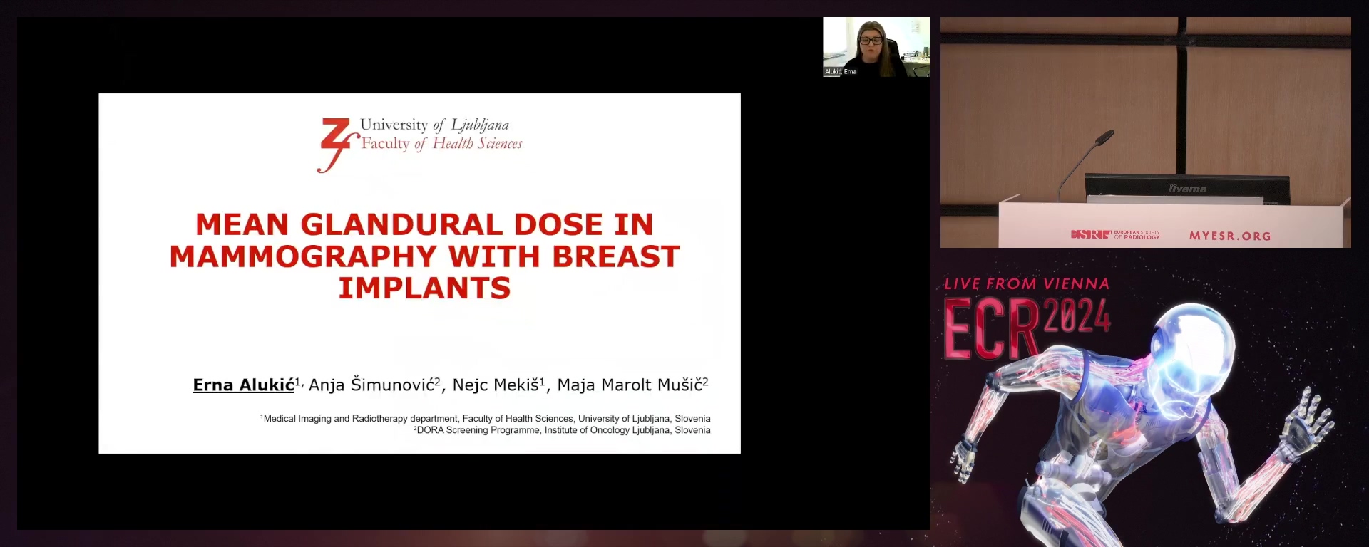
Mean glandular dose in mammography with breast implants
Erna Alukić, Ljubljana / Slovenia
- Author Block: E. Alukić, A. Simunovic, N. Mekis, M. Marolt Music; Ljubljana/SI
Purpose: The aim was to evaluate the mean glandular dose (MGD) in screening mammography in patients with breast implants and to investigate the differences in MGD depending on the type of exposure setting - automatic exposure control (AEC) or manual exposure technique.
Methods or Background: A retrospective study with secondary data analysis included 536 patients with breast implants who completed screening mammography between 2008 and
- MGD, breast thickness, and type of exposure setting were assessed and compared using standard mammography and the Eklund technique. Image quality was assessed by three radiologists experienced in mammography assessment. Results or Findings: MGD is statistically significantly lower for breast thicknesses of 6 < 8 cm, 8 < 10 cm, and > 10 cm for standard images with manual exposure technique. For additional images with the AEC system, the mean MGD is statistically significantly lower for breast thicknesses of < 4 cm and 4 < 6 cm. For additional images with breast thickness of 6 < 8 cm, the MGD value is significantly lower with the manual exposure technique. There are statistically significant differences in MGD between images taken with standard mammography and those taken with the Eklund technique for both types of exposure settings. The overall assessment of all quality criteria images was statistically significantly higher for the manual exposure technique. Conclusion: For additional images with breast thickness < 4 cm and 4 < 6 cm, the AEC system is better due to the lower MGD values. The manual mode is more suitable for breast thicknesses of 6 > 8 cm. Limitations: No limitations. Funding for this study: No funding was obtained for this study. Has your study been approved by an ethics committee? Yes Ethics committee - additional information: This study was ethically approved by the Commission of the Republic of Slovenia for Medical Ethics, Document number: ERIDEK-0047/
RPS 2214-10
7 min
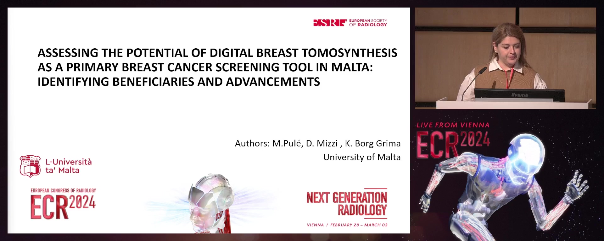
Assessing the potential of digital breast tomosynthesis as a primary breast cancer screening tool in Malta: identifying beneficiaries and advancements
Maria Pule, Paola / Malta
- Author Block: M. Pule1, D. Mizzi1, K. Borg Grima2; 1Msida/MT, 2Naxxar/MT
Purpose: The aim of the study was to prospectively identify who would benefit from having a digital breast tomosynthesis (DBT) as a first line screening. This was done by identifying different characteristics of patients being referred for a DBT at the local screening centre during further assessment clinics. The objectives of phase 1 were to audit the referrals for further assessment clinics within the local screening centre. In phase 2, data on the reason of referral together with the different patient characteristics, and the imaging results was collated to be able to reach set objectives.
Methods or Background: The study involved two sequential phases. Phase 1 consisted of a retrospective analysis of statistical figures on the use of DBT locally from 2015 to
- Phase 2 was a prospective study composed of a self-designed patient questionnaire distributed between March and April 2022, required to evaluate any link between the different patient characteristics and the clinical outcome following a DBT. Results or Findings: Phase 1 included 2,756 cases, where
- 9% had their first mammogram while 64.1% had a subsequent mammogram. First time mammogram cases were statistically the most likely to be returned back to normal screening (37.8% n=990). In both phases, asymmetric densities was the most common reason of referral. For phase 2, 53 participants were recruited. Results indicated that the most common types of Mammographic Breast Densities were type BIRADS A (30.2 %) and B (50.9%). Conclusion: From the results collected in both phases it was shown that women screened for the first-time would benefit from DBT as a first line screening tool since they are more likely to be returned back to normal screening. Limitations: Due to COVID-19 restrictions, the sample population for phase 2 was limited. Funding for this study: No funding was received for this study. Has your study been approved by an ethics committee? Yes Ethics committee - additional information: Approval for this research was then obtained from the Faculty Research Ethics Committee (FHS-2021-00023) and from the University Research Ethics Committee within the University of Malta.
