Research Presentation Session: Radiographers
RPS 1614 - Current insights and future horizons
RPS 1614-3
7 min
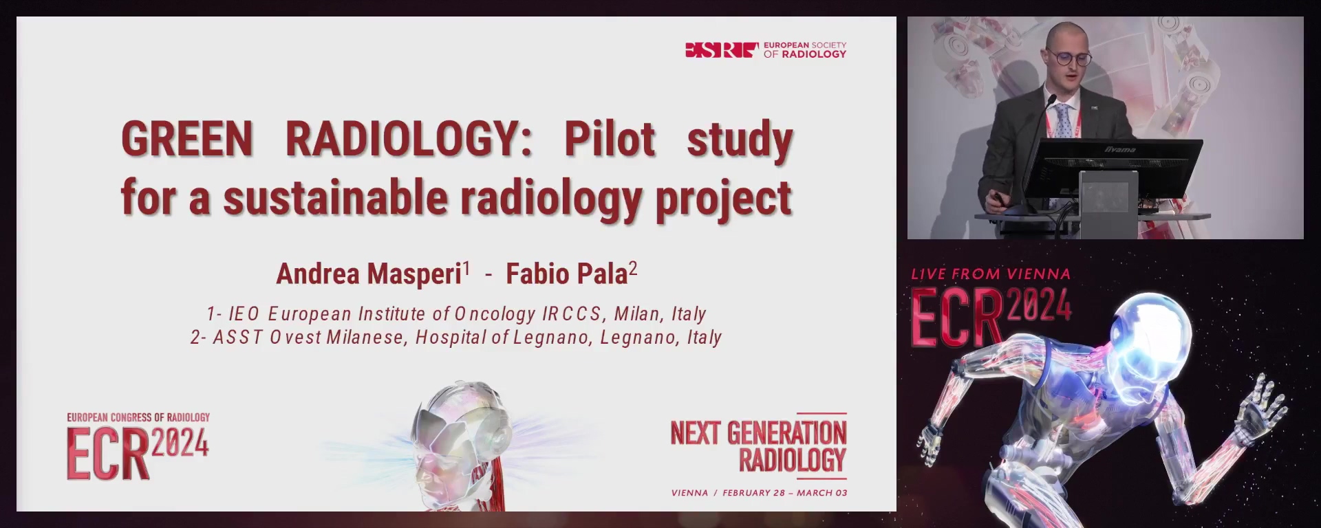
Green radiology: a pilot study for a sustainable radiology project
Andrea Masperi, Abbiategrasso / Italy
- Author Block: A. Masperi1, F. Pala2; 1Milan/IT, 2Legnano/ITPurpose: The purpose of this study was to estimate the energy impact of the radiology department, implement sustainable solutions in a testing period and evaluate the effects.Methods or Background: The project followed the energy diagnosis phases proposed by the UNI CEI/TR 11428 certification in a period between January and June
- A retrospective analysis mapped the activities and energy consumption of the functional categories involved imaging functional categories (IFC) and complementay functional categories (CFC) and 5 IFCs and 4 CFCs were defined as inefficient respectively. Four interventions were proposed such as optimisation of consumption, reallocation of diagnostic activities, awareness-raising in a green imaging protocol and staff training and a energy scorecard was drawn up. The proposed actions were deployed and the activities, energy consumption of critical IFCs and CFCs, and implementation of the green imaging protocol were monitored. Results or Findings: A reduction in total energy consumption of 8% was achieved for IFCs, and
- 2% for CFCs against an increase in average diagnostic activity of 11.8%. 41/156 patients (26.2%) were retrospectively evaluated with the green imaging protocol, recording a potential reduction in energy consumption of 378.1 kW/patient (77.3%). Conclusion: It is possible to identify with a fair degree of accuracy inefficient processes and functional devices that do not comply with the new guidelines proposed by the European Community for reducing energy consumption. Including radiographers in the drafting of energy dossiers offers the possibility of studying sustainable solutions that lead to concrete results, not only in terms of energy and money savings but also in terms of quality assistance.Limitations: We only considered electricity consumption and not energy consumption from other sources. The main limitations in terms of green protocol are attributable to the evaluation of simulated patients, within a limited period, limited to the abdominal study.Funding for this study: No funding was received for this study.Has your study been approved by an ethics committee? Not applicableEthics committee - additional information: No information provided by the submitter.
RPS 1614-4
7 min
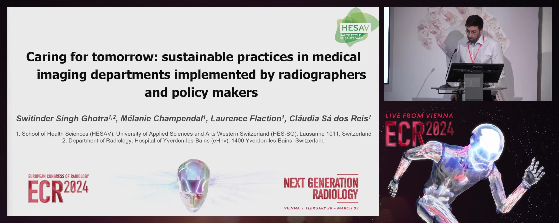
Caring for tomorrow: sustainable practices in medical imaging departments implemented by radiographers and policy makers
Switinder Singh Ghotra, Lausanne / Switzerland
- Author Block: S. S. Ghotra, M. Champendal, L. Flaction, C. S. D. Reis; Lausanne/CHPurpose: Global warming is one of the main public health concerns of our time. The purpose of this study was to identify salient approaches that can reduce the environmental impact of medical imaging departments/(MID).Methods or Background: A review was conducted following JBI methodology on PubMed, Embase and CINAHL to include studies published after 2013 (French, English). Combinations of keywords and MeSH terms related to environmental sustainability, recycling, medical waste and greening were applied. Three independent reviewers screened abstracts, titles and full text. Disagreements were solved through consensus.Results or Findings: 4630 studies were identified; 38 articles met all criteria. Most of the studies were related to developed countries (32/38) and 6/38 were from non-developed countries. A third of the studies included were published after
- Articles focused on computed tomography (9/38), magnetic resonance imaging (6/38), interventional radiology (4/38), conventional radiography (4/38), ultrasound (2/38), mixed modalities (9/38). Seven main categories to reduce environmental impact were identified: 1) examination justification, 2) energy consumption, 3) waste production, 4) recycling opportunities, 5) local resources usage, 6) environmental pollution and 7) education. The study indicates the salience of sustainability analysis within quality assurance programmes. Conclusion: To reduce the environmental impact of MID it is important to educate healthcare professionals and to justify adequately examinations, to control energy consumption and to improve health outcomes. Further studies need to be conducted to identify strategies that are most effective, supporting the decision making of managers and MI professionals.Limitations: No experimental studies were conducted to identify the most cost-effective strategies.Funding for this study: No funding was received for this study.Has your study been approved by an ethics committee? Not applicableEthics committee - additional information: No information provided by the submitter.
RPS 1614-5
7 min
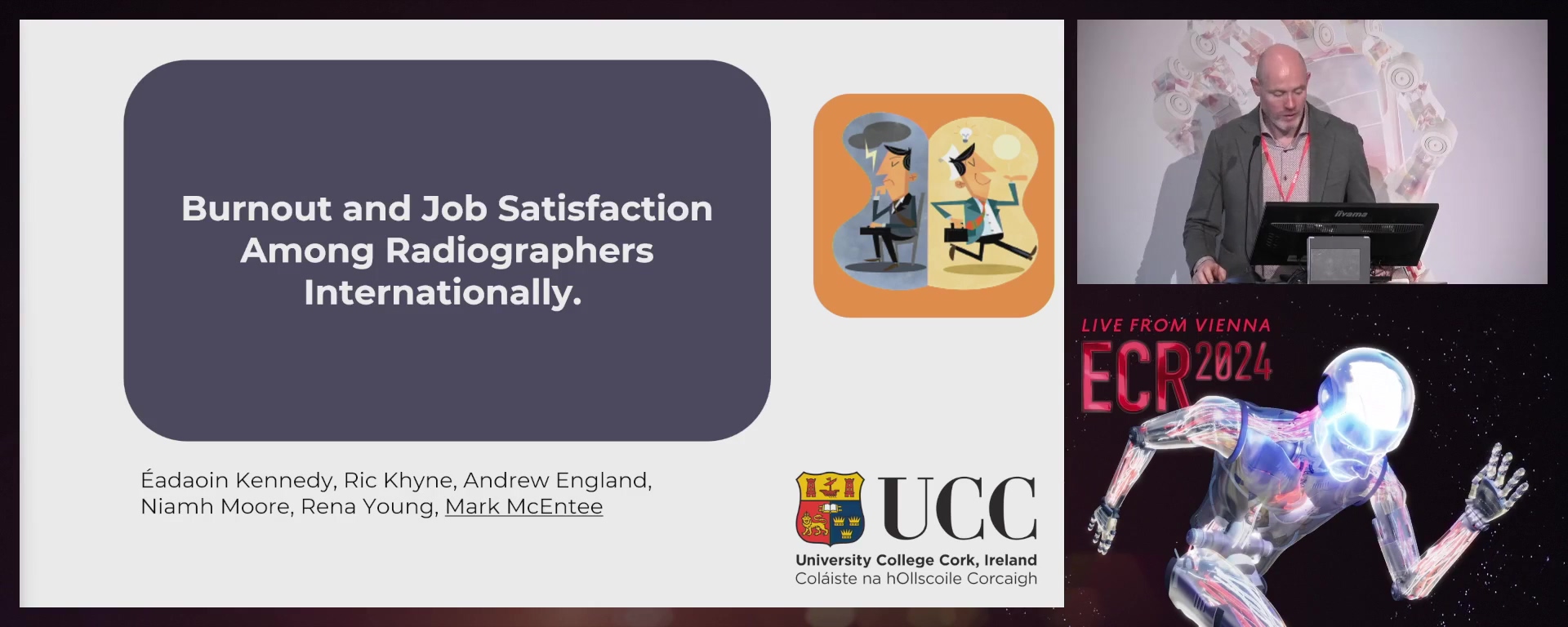
Burnout and job satisfaction among radiographers internationally
Mark F. McEntee, Cork / Ireland
- Author Block: E. Kennedy, A. England, N. Moore, R. Young, M. F. F. McEntee; Cork/IEPurpose: Burnout and low job satisfaction in healthcare can impact patient safety and staff retention. This study aims to gain information on the factors influencing the levels of burnout and job satisfaction among radiographers internationally, which can inform strategies for improving workforce supply and demand imbalance.Methods or Background: An online questionnaire was developed that included demographic questions and two validated instruments, The Maslach Burnout Inventory (MBI) and the Job Satisfaction Survey (JSS). Statistical analysis was performed using IBM SPSS. The questionnaire was distributed to state registered diagnostic radiographers through the EFRS Research Hub in 2023 and online through twitter, facebook and e-mail over a six-week period.Results or Findings: 247 participants completed the questionnaire and 207 (
- 5%) were female. The questionnaire had participants from 21 countries, with 66.5% being from Ireland. The mean values for EE (20), DP (11) and PA (36) indicate moderate levels of burnout among responding radiographers. 44.2% of radiographers were dissatisfied, 43.7% were ambivalent and only 12.1% had overall job satisfaction. Workload, work being underappreciated, and time pressures were ranked as the top three factors contributing to burnout. Staff numbers, workload and poor management, were ranked as the top three factors reducing job satisfaction. Conclusion: Burnout levels were moderate and overall job satisfaction was very low in participating radiographers. Workload and being underappreciated were identified among many factors that impact job satisfaction and burnout.Limitations: Despite the international intent of the study, two thirds of participants were from the Republic of Ireland.Funding for this study: No funding was received for this study.Has your study been approved by an ethics committee? YesEthics committee - additional information: This study was approved by the Medical School SREC, University College Cork.
RPS 1614-6
7 min
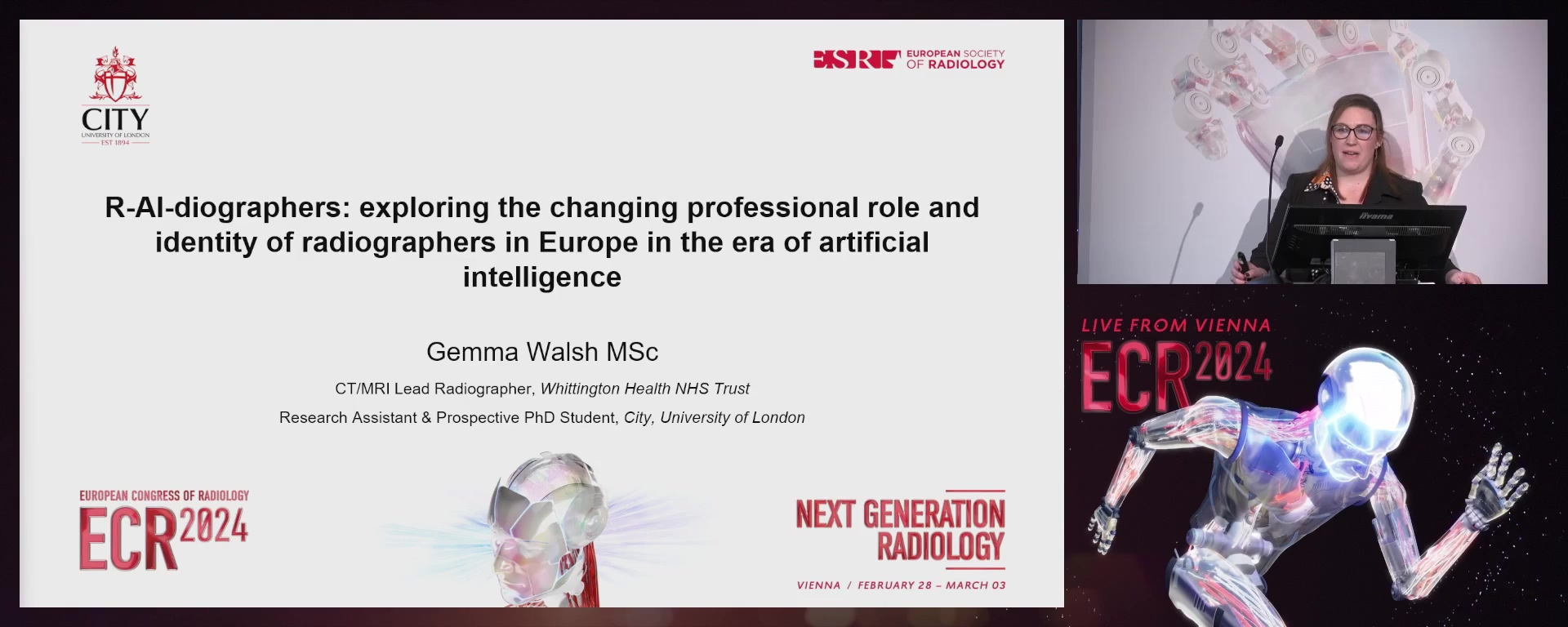
R-AI-diographers: exploring the changing professional role and identity of radiographers in Europe in the era of artificial intelligence (AI)
Gemma Walsh, Chesham / United Kingdom
Author Block: G. Walsh1, M. F. F. McEntee2, Y. Kyratsis3, C. A. Beardmore1, C. Malamateniou1; 1London/UK, 2Cork/IE, 3Amsterdam/NL
Purpose: This study aims to gain insights into the changing roles and identities of diagnostic and therapeutic radiographers in the era of AI. The objective is to propose ways to better support the workforce in the face of fast technological changes.
Methods or Background: Ethics approval and written informed consent was gained prior to data collection. A Europe-wide, cross-sectional study utilising a mixed methods online survey was designed with key stakeholder feedback and translated from English into eight languages. Snowball sampling was used for distribution via social media. All European radiographers (including students) were eligible to participate. The survey collected data on the following areas: a) demographics, b) the perceived short-term impact of AI on radiographer roles, c) the potential medium-to-long-term impact of AI, d) perceived opportunities and threats of AI implementation for radiographers' roles and careers, e) the preparedness of radiographers to work with AI and f) the potential for future AI leadership roles for radiographers.
Results or Findings: A total of 2,258 valid responses from 38 European countries were received. Country of practice, gender, modality expertise, and years of experience impacted the responses. Training quality and quantity influence the perceptions of AI. Despite some concerns around job security, survey responses were collectively projecting a feeling of optimism for the future of radiographer careers and professional identity. Knowledge, additional training, job satisfaction, better patient care, financial compensation and career advancement were presented as key motivators to professional engagement with AI.
Conclusion: This study provides insights into radiographers' attitudes towards AI implementation on the future of their professional identity. It proposes ways to better support the workforce in harnessing the benefits of AI.
Limitations: Survey translators were not professional translators but radiographers with expert knowledge of subject terminology.
Funding for this study: CoRIPS from the College of Radiographers and City Radiography Research Fund.
Has your study been approved by an ethics committee? Yes
Ethics committee - additional information: This study was approved by the ethics committee of City, University of London.
RPS 1614-7
7 min
What radiographers need to know about explainable artificial intelligence in medical imaging?
Mélanie Champendal, Lausanne / Switzerland
- Author Block: M. Champendal1, H. Müller2, J. O. Prior1, C. S. D. Reis1; 1Lausanne/CH, 2Sierre/CHPurpose: Artificial Intelligence/(AI) is seen as a "black box" and health professionals tend to not trust it, at least not fully. This study aimed to present to radiographers the main eXplainable Artificial Intelligence/(XAI) methods available for medical imaging/(MI) allowing them to understand it.Methods or Background: A review was conducted following JBI methodology, searching PubMed, Embase, Cinhal, Web of Science, BioRxiv, MedRxiv, Google Scholar for French and English studies post 2017 on explainability and MI modalities. Two reviewers screened abstracts, titles, full texts, resolving disagreements through consensus.Results or Findings: 1258 results were identified; 228 studies meet all criteria. The terminology used across the articles varied: explainable (n=207), interpretable (n=187), understandable (n=112), transparent (n=61), reliable (n=31), intelligible (n=3) being used interchangeably. XAI tools applied to MI are mainly intended for MRI, CT and x-ray imaging to explain lung/(Covid) (n=82) and brain/(Alzheimer's; tumours) (n=74) pathologies. The main formats used to explain the AI tools were visual (n=186), numerical (n=67), rule-based (n=11), textual (n=11). The classification (n=90), prediction (n=47), diagnosis (n=39), detection (n=29), segmentation (n=13) and image quality improvement (n=6) were the main tasks explained. The most frequently provided explanations were local (
- 1%); 5.7% were global, and 16.2% combined both local and global approaches. Conclusion: The number of XAI publications in MI is increasing, in aid of the classification and prediction of lung and brain pathologies. Visual and numerical output formats are predominantly used. Terminology standardisation remains a challenge, as terms like "explainable" and "interpretable" are used as equivalent. Future work is required to integrate all stakeholders in the construction of XAI.Limitations: The focus was solely on recent XAI developments, leading to the exclusion of studies published before 2018, which may have caused other tools that were explored during that period to be excluded.Funding for this study: No funding was received for this study.Has your study been approved by an ethics committee? Not applicableEthics committee - additional information: No information provided by the submitter.
RPS 1614-8
7 min
Identifying the needs of radiographers working in nuclear medicine departments for the co-construction of XAI tools for enhanced PET image reconstruction
Mélanie Champendal, Lausanne / Switzerland
Author Block: M. Champendal1, H. Müller2, J. O. Prior1, C. S. D. Reis1; 1Lausanne/CH, 2Sierre/CH
Purpose: AI is often viewed as a 'black box,' causing some distrust among healthcare professionals. This study aimed to pinpoint the needs of radiographers working in a nuclear medicine department to understand better the AI algorithm used for enhancing PET/CT images.
Methods or Background: Two Focus Groups/(FG) were conducted to identify background knowledge and perspectives about AI. An introduction about XAI was performed, and specific needs and preferences were explored through the presentation of scenarios, giving results corresponding or not corresponding to the ground truth. The questions that XAI should explain to support radiographers in their practice were collected, as well as main characteristics in terms of output format, confidence levels, barriers and facilitators for AI use. Thematic analysis was carried out following the Braun & Clark Framework.
Results or Findings: Ten radiographers (aged 31-60) from various hospital settings discussed their needs for XAI. While three currently use AI tools in PET/CT with limited training, their main requirements for XAI tools include interactivity, adaptability, user-friendliness, and minimal workflow disruption. They found a 'trust index,' visual comparisons, example-based, and chatbots as valuable output formats. Facilitators for XAI use included training, support, early integration, and radiographers specialists, while barriers included lack of understanding, organisational challenges, and system capabilities.
Conclusion: Needs for XAI tools were identified, but it is important to improve radiographers' knowledge and prepare the system to implement it without negatively impacting the workflow and patient outcomes.
Limitations: Limitations were the focus group sample size impacting generalisation and XAI knowledge.
Funding for this study: No funding was received for this study.
Has your study been approved by an ethics committee? Not applicable
Ethics committee - additional information: This study did not require ethics committee approval since no personal data were collected.
RPS 1614-12
7 min
The role of virtual reality allowing dialogue between users and virtual patients for the preclinical education of radiographers: a pilot study
Kengo Kato, Tsukuba / Japan
- Author Block: K. Kato1, D. Kon2, K. Ueda2, C. S. D. Reis3, N. Ienaga1, Y. Kuroda1; 1Tsukuba/JP, 2Narita/JP, 3Lausanne/CHPurpose: Head-mounted display-based immersive virtual reality (VR) coaching systems (HMD-VRC) in radiographers' training often lack adequate interaction through spoken dialogue between users and virtual patients (VP). To enable comprehensive training, including the confirmation process to prevent risks, the study aimed to develop an HMD-VRC that allows interaction with VP to enhance preclinical learning.Methods or Background: HMD-VRC replicates 3D models of an x-ray room and the VP, incorporating two main features: 1) a voice bot based on Dialogflow, Google LLC, for the VP to respond to user dialogue and 2) an on-screen hint display system for learning support. As a pilot study, first-year students (n=10) from a Radiography/Medical Imaging (MI) school without specialised education were given a 20-minute explanation using text and physical equipment. Subsequently, they received radiography education using HMD-VRC for approximately one hour. Objective assessments via role-playing with actual equipment were conducted before and after HMD-VRC education. A survey regarding HMD-VRC was administered, asking about the experience and learning process.Results or Findings: HMD-VRC enabled all students to complete radiography training from patient entry to exit. Through HMD-VRC training, the average score of the objective assessments with actual equipment increased from
- 2% to 88.3%, nearly 1.5 times higher. Survey results suggested a deeper understanding of the process through HMD-VRC, highlighting the importance of communicating/discussing with the VP in training. Additionally, HMD-VRC training was found to alleviate anxiety in objective assessments via role-playing. Conclusion: This pilot study suggests that education using HMD-VRC with responsive VP through spoken dialogue is effective for preclinical education, promoting practical learning for radiographers' training. This innovative approach holds promise for improving Radiography/MI training programs, facilitating the transition between theory and practice.Limitations: Only chest radiography was considered, other body parts will be validated.Funding for this study: This study was partly supported by the JSPS KAKENHI (JP23K11369).Has your study been approved by an ethics committee? YesEthics committee - additional information: This study was approved by the ethics committee of the International University of Health and Welfare (Approval No. 23-Im-030).
RPS 1614-13
7 min
The inclusion of cross-sectional imaging within preregistration radiography training curricula
Rena Young, Cork / Ireland
Author Block: M. Friel, R. Young, N. Moore, M. F. F. McEntee, A. England; Cork/IE
Purpose: Clinical practice is a critical component of all radiography curricula. It provides students with essential skills and training, enabling them to develop professional characteristics for competent radiography practice. Educational institutions are challenged with regularly reviewing and developing strategies to ensure that curricula satisfy evolving service needs. This study aimed to gain insight into the inclusion of cross-sectional imaging (CT/MRI) within preregistration radiography training curricula.
Methods or Background: An online questionnaire, based on previous EFRS surveys, included open and closed-ended questions, and ascertained on level of qualification, cross-sectional imaging incorporation, and tasks and assessments within programmes. The questionnaire was distributed through the EFRS Research Hub at the European Congress of Radiology (ECR2023), and online via social media. Closed questions were summarised using descriptive statistics, while open-ended questions were thematically analysed.
Results or Findings: Responses were received from 64 radiography educational institutions within 29 different countries. The inclusion of cross-sectional (CT/MRI) training was reported by 57 respondents (92%) as part of their institution's radiography programme. An increase in the amount of time dedicated to clinical training in cross-sectional imaging was reported by 24 respondents (38%). Overall, 32 individuals (53%) stated that CT should be reserved as a specialised modality for radiographers once they attain their basic qualification, while 36 respondents (60%) agreed that MRI should also be reserved as a specialised modality.
Conclusion: The findings indicate a lack of uniformity among preregistration radiography programmes, regarding cross-sectional imaging incorporation within their curricula. However, there appear to be divided views on the issue.
Limitations: Predominantly, responses to the survey came from countries where English is the first/dominant language.
Funding for this study: No funding was received for this study.
Has your study been approved by an ethics committee? Yes
Ethics committee - additional information: This study was approved by the Medical School SREC, University College Cork.
RPS 1614-14
7 min
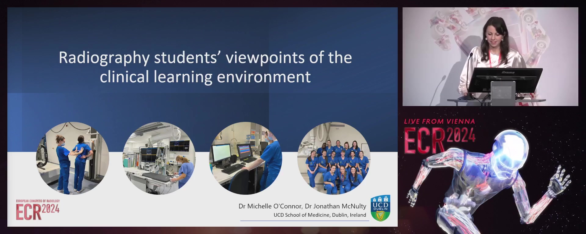
Radiography students' viewpoints of the clinical learning environment: a cross-sectional study
Michelle O'Connor, Dublin / Ireland
- Author Block: M. O'Connor, J. McNulty; Dublin/IEPurpose: Clinical practice is fundamental to the development of students' competence and professionalism. Objective instruments have been developed to monitor the quality of clinical learning environments (CLEs) in medicine and nursing. This study is the first to investigate radiography students' perspectives of their CLEs using the validated Undergraduate Clinical Education Environment Measure (UCEEM).Methods or Background: Undergraduate (UG) (n=365) and graduate-entry (GE) radiography students (n=45) from an Irish university were invited to complete the online, self-administered, questionnaire regarding their recent clinical placement. CLEs consisted of 25 public and 10 private hospitals nationally. The UCEEM contains 25 items congregated into two overarching dimensions, experiential learning and social participation, with four subscales: opportunities to learn in and through work and quality of supervision; preparedness for student entry; workplace interaction patterns and student inclusion; and equal treatment.Results or Findings: 215 students participated (response rate
- 4%) (n=185 UG, n=30 GE), most of whom were based in public hospitals (87.4%). The mean total UCEEM score was 107.54±17.67 (optimal range: 90–125). Mean scores for 'experiential learning and social participation' were 74.2±12.5 (optimal range: 60–85) and 33.37±6.29 (optimal range: 30-40), respectively. Private hospitals ranked slightly higher than public hospitals for 'opportunities to learn in and through work and the quality of supervision' (p <0.05). The highest ranked items related to 'equal treatment' and the 'opportunity to put theory into practice'. Few statistically significant differences existed between cohorts except for clinical supervision-related items which were scored highest by first year students. Conclusion: Students across both programmes reported high levels of satisfaction with their CLEs in terms of experiential learning and social participation. This study provides valuable baseline data for comparison of Radiography CLEs.Limitations: The study was limited to a single Irish university.Funding for this study: No funding was received for this study.Has your study been approved by an ethics committee? YesEthics committee - additional information: The study was approved by University College Dublin (LS-C-22-153-Oconnor).
