Research Presentation Session: Radiographers
RPS 1014 - Optimisation of practice, care and safety: tips and tricks
RPS 1014-3
7 min
A phantom image quality study comparing ultra-low and standard dose computed tomography protocols for the investigation of non-accidental injury
Niamh Moore, Clogheen / Ireland
- Author Block: L. Kingston, M. F. F. McEntee, N. Moore, R. Young, C. S. Lee, A. England; Cork/IEPurpose: The purpose of this study was to determine if there is potential for the development and utilisation of a low dose computed tomography (CT) protocol for the diagnosis of suspected non-accidental injury (NAI) and to establish if the suggested ultra-low dose (ULD) CT protocol could deliver image quality (IQ) comparable to that of a standard dose (STD) CT protocol.Methods or Background: CT images were acquired on a Revolution Apex™ scanner, using a new-born whole body phantom, and reconstructed using deep learning software. Two protocols were compared in terms of IQ, an ULD protocol, with a dose-length product (DLP) of
- 49mGycm2 and a STD protocol, with a DLP of 22.92mGycm2. Participants graded IQ using a modified 4-point Likert scale. Mann-Whitney and chi-squared tests were carried out to determine if there was a significant difference in IQ between the protocols. A t-test was performed to determine if there was a significant difference in participants level confidence in the IQ between the protocols. Results or Findings: 27 participants met the inclusion criteria and scored both image sets. Both the Mann-Whitney and chi-squared test demonstrated significant difference (P ≤
- 05) in the IQ between the protocols, for both the evaluation of cortical and trabecular bone. T-test results demonstrated a significant difference (P ≤ 0.05) in the level of confidence amongst participants between the protocols. Conclusion: This phantom-based study suggests that the IQ of the STD protocol was significantly better when compared to the ULD protocol, for the investigation of suspected NAI. Further work is required to fully optimise an ULD-CT protocol for the investigation of NAI.Limitations: This was a phantom-based study using images without the presence of pathology.Funding for this study: No funding was received for this study.Has your study been approved by an ethics committee? YesEthics committee - additional information: This study was approved by the Medical School SREC, University College Cork.
RPS 1014-4
7 min
Evaluation of anatomical structures by pulmonary angio-CT in patients with PTE and COVID-19
Ana Filipa Colucas, Olhão / Portugal
- Author Block: A. F. Colucas, S. I. Rodrigues, L. P. V. Ribeiro, A. F. C. L. Abrantes, O. Lesyuk; Faro/PTPurpose: The aim of this study was to identify anatomical changes in the pulmonary angio-CT associated with the clinical worsening of COVID-19 and pulmonary thromboembolism. Results were compared between non-COVID-19 patients and COVID-19 patients, in order to understand whether the presence of this infection interfered with the workflow at the hospital level and whether it had an influence on changes in anatomical structures at the cardiac level.Methods or Background: This quantitative descriptive-correlational study with a sample of 134 angio-CT exams was conducted in a public hospital, on patients with pulmonary Thromboembolism (PTE), separated into two groups; 81 non-Covid-19 patients and 53 COVID-19 patients. Measurements were taken of the Right Ventricle (RV), Left Ventricle (LV) and Pulmonary Artery Trunk (PAT) to assess whether there was RV dilatation (when RV/LV >1).Results or Findings: Pulmonary angio-CT scans have increased 88% since the COVID-19 pandemic and consequently, an increase in positive diagnoses for PTE. Regarding the results,
- 2% and 82.7% of the non-COVID-19 patients showed dilatation of the RV and dilatation of the PAT respectively. Considering the COVID-19 group, 92.5% and 96,2% of the patients showed dilatation of the RV and PAT, respectively. Considering the Spearman test, the correlation between "COVID-19" and the "RV dilatation" is verified. Conclusion: Comparing the results for both groups, it was found that the group with COVID-19 presented a higher incidence of anatomical changes at the cardiac level, and male gender has been a predictive factor for the presence of both pathologies.Limitations: It is difficult to identify if the non-covid patients had previous infections and a bigger sample is necessary to evaluate the changes in anatomical structures.Funding for this study: No funding was received for this study.Has your study been approved by an ethics committee? YesEthics committee - additional information: This study was approved by the ethics committee; decision number 164/
RPS 1014-5
7 min
Ultra-low-dose CT protocol for left ventricular ejection fraction (LVEF) and combination with chest-abdomen-pelvis CT
Martin Weber Kusk, Esbjerg / Denmark
- Author Block: M. W. Kusk1, S. Hess2, O. Gerke2, C. Stolzenburg Oxlund1, L. Deibjerg Kristensen1, T. E. J. Ormstrup1, J. M. Christiansen1, S. J. Foley3; 1Esbjerg/DK, 2Odense/DK, 3Dublin/IEPurpose: The aim of this study was to test the accuracy of ultra-low-dose CT for left ventricular ejection fraction (LVEF-CT). A secondary objective was to examine the feasibility of combination with routine Chest-Abdomen-Pelvis (CAP) CT in oncology patients as potential one-stop replacement for nuclear multigated (MUGA) scans to monitor chemotherapy cardiotoxicity.Methods or Background: Patients underwent LVEF-CT with cardiac MRI as reference. In oncology patients, MUGA scans were also performed and LVEF-CT was combined with CAP CT, with modified injection protocol. For each modality, two readers measured LVEF. We assessed bias using a Bland-Altman analysis and correlation via Pearson correlation. Interreader agreement was measured using ICC. ROC analysis was performed using MRI 50% LVEF cut-off for reduced LVEF and sensitivity/specificity calculated at maximum Youden Index. We compared CAP-scans to previous scans, using ROI mesasurements, visual grading characteristics and diagnostic acceptability scores.Results or Findings: 77 datasets of 82 patients were suitable. Mean doses were
- 4 mSv for LVEF-CT, 5.7 mSv for MUGA and 7.4 mSv for CAP. CT-derived LVEF bias to MRI varied from 2-10 %, dependent on measurement modes. ROC AUCs exceeded 0.95 and sensitivity/specificity of 100% were achieved with cut-offs from 53-63% LVEF. CT-derived LVEF correlated better with MRI (R=0.87 vs 0.71) and had higher ICC than MUGA (0.98 vs 0.62). CAP image quality showed decreased renal medulla/cortex discrimination, but the number of diagnostic scans was not significantly different from previous scans. Mean examination times were 16, 28, and 32 minutes for CT, MRI and MUGA, respectively. Conclusion: CT LVEF measurement is feasible at a quarter of MUGA dose and can be combined with chest-abdomen-pelvis CT, for a one-stop examination.Limitations: Temporal changes in LVEF were not examined. MUGA subgroup had limited patients with reduced LVEF to measure MUGA classification accuracy.Funding for this study: The study was funded by the Esbjerg Fund (IC Møllers Fond), the research fund of the Danish Radiographers Association (Radiograf Rådet) and the Karola Jørgensen Fund for health research at Esbjerg University Hospital.Has your study been approved by an ethics committee? YesEthics committee - additional information: This study was approved by the regional committees on health research ethics for Southern Denmark (approvals. no. S-20210094 and S-20220031).
RPS 1014-6
7 min
Radiomic features of kidney stones with use of virtual non-contrast on photon counting CT: a phantom study
Simon Lysdahlgaard, Esbjerg / Denmark
- Author Block: S. Lysdahlgaard1, L. N. Hansen1, I. Riebenholt Nielsen1, B. R. Mussmann2, J. Jensen2, H. U. Jung3, M. W. Kusk1; 1Esbjerg/DK, 2Odense/DK, 3Vejle/DKPurpose: This study aimed to assess the accuracy of kidney stone radiomics and Hounsfield unit (HU) measurements using the Virtual Non-contrast (VNC) technique compared to the traditional True Non-contrast (TNC).Methods or Background: A Gammex RMI-461a head/body phantom incorporating 18 brushite and 18 uric acid kidney stones. All stones were scanned using low-dose non-contrast computed tomography (NCCT) and standard-dose abdominal protocols. Scans were conducted both without and with stones embedded in contrast media. Images were reconstructed using the VNC technique. Each stone was segmented using 3D slicer and
- 150 radiomics features were extracted. T-tests were used to test for statistically significant differences in mean feature values between stone classifications, with Bonferroni correction applied. A p-value of > 0.05 was considered significant. Results or Findings: Radiomic features from brushite and uric acid for abdomen and NCCT protocols with contrast media yielded 286 and 285 significant values (p <
- 05), respectively. Following Bonferroni correction, these numbers were 35 and 31 values (p < 0.000044). Radiomic features for both stone types using abdomen and NCCT protocols without contrast media were 446 and 466 significant values (p < 0.05), with Bonferroni correction yielding 238 and 224 values (p < 0.000044), respectively. Comparative analysis showed that 17 radiomic features were significant in scans in TNC, while 196 features were significant in scans in VNC. Conclusion: This study found fewer statistically significant radiomic features between brushite and uric acid stones in VNC compared to TNC, for both NCCT and abdomen protocols scanned on PCCT. Radiomic features extracted from different stone types further emphasise the potential of this method in enhancing diagnostic precision.Limitations: This study was a phantom study, with only two types of kidney stones, and an operator-dependent segmentation tool.Funding for this study: No funding was received for this study.Has your study been approved by an ethics committee? Not applicableEthics committee - additional information: Consent was waived by the ethics committee.
RPS 1014-7
7 min
An investigation into the impact of radiation dose information on the subjective evaluation of CT image quality
Rena Young, Cork / Ireland
Author Block: C. S. Lee, L. Kingston, M. F. F. McEntee, R. Young, N. Moore, M. Maher, A. England; Cork/IE
Purpose: The use of medical imaging radiation exposure has dramatically increased in recent years and is continuing to do so. Ultra-low dose examinations may combat this, but switching to ultra-low dose protocols in CT is difficult due to the potential loss of image quality. This has been assessed within numerous studies which have found high interobserver variability. One possible reason for this high variability is the presence of contextual bias. This study aims to investigate the presence and magnitude of bias created through the presence of radiation dose information on images.
Methods or Background: Two CT datasets of images were created using a paediatric phantom. The phantom was scanned using a 'standard' dose protocol and an 'ultra-low' dose protocol. Participant 'radiographers' were asked to score the image quality of each image using the 'PGMI' scoring system. Participants were requested to focus on scoring image quality for cortical and trabecular bone, across the full skeleton. Participants were given the images either with or without the dose information present. Data was analysed statistically.
Results or Findings: 53 participants were included. 25 completed the dataset with the dose information present, and 38 completed the dataset without the dose information. Overall, it was found that the dataset with dose information provided received a 23% and 22% higher score for both cortical and trabecular bone image quality, respectively for the standard dose images and 7% and 4% for those acquired using the ultra-low dose protocol.
Conclusion: The addition of radiation dose information created contextual bias in both datasets and it had a larger impact on the standard dose CT images.
Limitations: This study was based on CT images acquired using a paediatric phantom.
Funding for this study: No funding was received for this study.
Has your study been approved by an ethics committee? Yes
Ethics committee - additional information: This study was approved by the Medical School SREC, University College Cork.
RPS 1014-8
7 min
Implementation and standardisation of CT optimisation tools across Europe: results from the iViolin project survey
Nadin Jamal Abualroos, Dublin / Ireland
- Author Block: S. J. Foley, J. McNulty, N. J. Abualroos; Dublin/IEPurpose: To identify the current availability and implementation of CT optimisation tools during lung, colorectal, and stomach cancer routine scanning protocols across different centres in Europe.Methods or Background: An online survey consisting of 18 questions was disseminated to six different clinical sites across Europe via iViolin management team in January
- The survey was addressed to key imaging representatives (namely a radiologist, radiographer and medical physicist) from each of the six contributing university hospitals. The survey analysed the availability and frequency of use of the available optimisation tools during lung, colorectal, and stomach cancer routine scanning protocols. Results or Findings: Responses were received from all six centres in five European countries with data reported for 22 CT scanners (Siemens: 64%, Philips: 18%, GE: 14%, Canon: 5%). Significant variations in the availability and utilisation of CT optimisation tools in different centres were identified, with automatic exposure control and iterative reconstruction algorithms widely available and in frequent use. The least available tools were additional filtration, high-pitch scanning, and deep-learning reconstruction. Respondents stated that barriers to implementation include subjective local preferences.Conclusion: Results demonstrate varying availability and use of CT optimisation tools. While surveyed centres have access to many optimisation tools, some challenges remain in their full implementation and usage.Limitations: Primary limitation is the limited sample size consisting of just six hospitals from five countries but convenience sampling was used to expedite data collection.Funding for this study: This project is co-funded under the EU4Health Programme 2021–2027 under grant agreement no. Has your study been approved by an ethics committee? Not applicableEthics committee - additional information: As a low risk study which collected data on CT protocols in current use and information was not sensitive application for full ethical review was not necessary
RPS 1014-9
7 min
Evaluation of sexual dimorphism of paranasal sinus and sella turcica by computed tomography: a retrospective study of a Portuguese population
Rosa Ramos Gaspar, Coimbra / Portugal
- Author Block: R. R. Gaspar1, M. Lopes2, T. Ribeiro2, B. M. Magalhães1, P. M. Martins2; 1Coimbra/PT, 2Aveiro/PTPurpose: A potential for sexual dimorphism has been pointed out regarding the study of paranasal sinuses (PS) and sella turcica (ST) in a forensic context, but results are conflicting in the literature. The aim of this study is to ascertain if PS: maxillary sinus (MS) sphenoidal sinus (SS) and ST measurements present sexual dimorphism in a Portuguese population.Methods or Background: This was a retrospective, observational study including 53 CT scans of the PS from Portuguese subjects (31 males/22 females. Exclusion criteria were age below 18 years, trauma, tumours, previous surgery, and sinus dysplasia. Measurements of length (AP), width (WI), height (HE), distance between MS (BIL), and volume of MS were obtained bilaterally; for the SS AP, WI, and HE were also obtained. ST measures included AP diameter, depth, and length. All linear measurements were done with RadiAnt DICOM Viewer. The volume of the MS was estimated from linear measurements. Data were analysed using SPSS-
- Normality and homogeneity were assessed. T-student and Mann-Whitney tests were applied. Statistical significance was set at .05. Results or Findings: Males present mean values higher than females except for measures of the sella turcica, in all instances. Statistically significant differences between sexes were found for five of the measurements: left WI (p=.042), left HE (p=.004), left volume (p=.033) of the MS, HE of the SS (p=.011), and ST AP Diameter (p=.016). A discriminant equation involving the measures with normal distribution was done. The height of the left MS shows an accuracy of 66% in predicting sex.Conclusion: The present study reveals that, at least, the height of the maxillary sinus presents sexual dimorphism. The methods hold promise in the field of forensic sciences to characterise the Portuguese population.Limitations: A larger sample size should be considered.Funding for this study: No funding was received for this study.Has your study been approved by an ethics committee? Not applicableEthics committee - additional information: In this retrospective study, the data under analysis were subject to irreversible anonymisation and stored by the investigator in an encrypted digital format during this research.
RPS 1014-10
7 min
The application of low-dose chest CT for the diagnosis and monitoring of pulmonary infections in neutropenic patients
Efthimios M. Agadakos, Athens / Greece
- Author Block: E. M. Agadakos, A. Zormpala, N. Zaios, C. Kapsiocha, M. Voulgarelis, N. Sypsas, L. A. Moulopoulos, V. Koutoulidis; Athens/GRPurpose: The aim of our study was to investigate the image quality and the diagnostic performance of low-dose Chest CT (LDCCT) for the diagnosis and monitoring of pulmonary infections in neutropenic patients.Methods or Background: Data from 164 consecutive neutropenic patients of a total of 256 CT examinations [149 LDCCT and 107 Standard Dose Chest CT (SDCCT)] were obtained between May 2015 and June 2019 . Examiners analysed the image quality and evaluated the radiologic findings associated with pulmonary infections to determine differences in diagnostic performance between the two imaging protocols.Results or Findings: The LDCCT protocol reduced radiation dose to patients by 47%. LDCCT images compared well in terms of image noise and image quality to SDCCT images. Detection of consolidation and ground glass opacity was significantly lower with the LDCCT protocol (LDCCT:
- 5% and 64.4%, respectively, SDCCT: 45.8% and 82.2%, respectively) with all the respective p-values, unadjusted and adjusted for sex, age and BMI analyses as well as the corresponding odds ratios. Similarly, LDCCT diagnostic sensitivity rated lower for nodules ≥3mm and ground glass halo in nodule(s) but was not affected by sex, age and BMI. There were no statistically significant differences in diagnostic performance between the two protocols for the detection of cavitation in nodules, diffuse interlobular septal thickening, pleural effusion, pericardial effusion and lymphadenopathy. Conclusion: LDCCT achieved a remarkable dose reduction, image quality and noise levels rated well yet underestimated four important radiologic findings associated with pulmonary infections in neutropenic patients.Limitations: The limitations encountered during the current study were the lack of follow-up of patient progress and the effect on the prognosis and survival rate. Another limitation was that both data sets were obtained using only a single IR algorithm strength setting.Funding for this study: This study did not receive any specific grant from funding agencies in the public, commercial, or not-for-profit sectors.Has your study been approved by an ethics committee? YesEthics committee - additional information: The study was compliant with the General Data Protection Regulation (679/2016 EE) and was approved by the hospital's scientific council and ethics committee.
RPS 1014-11
7 min
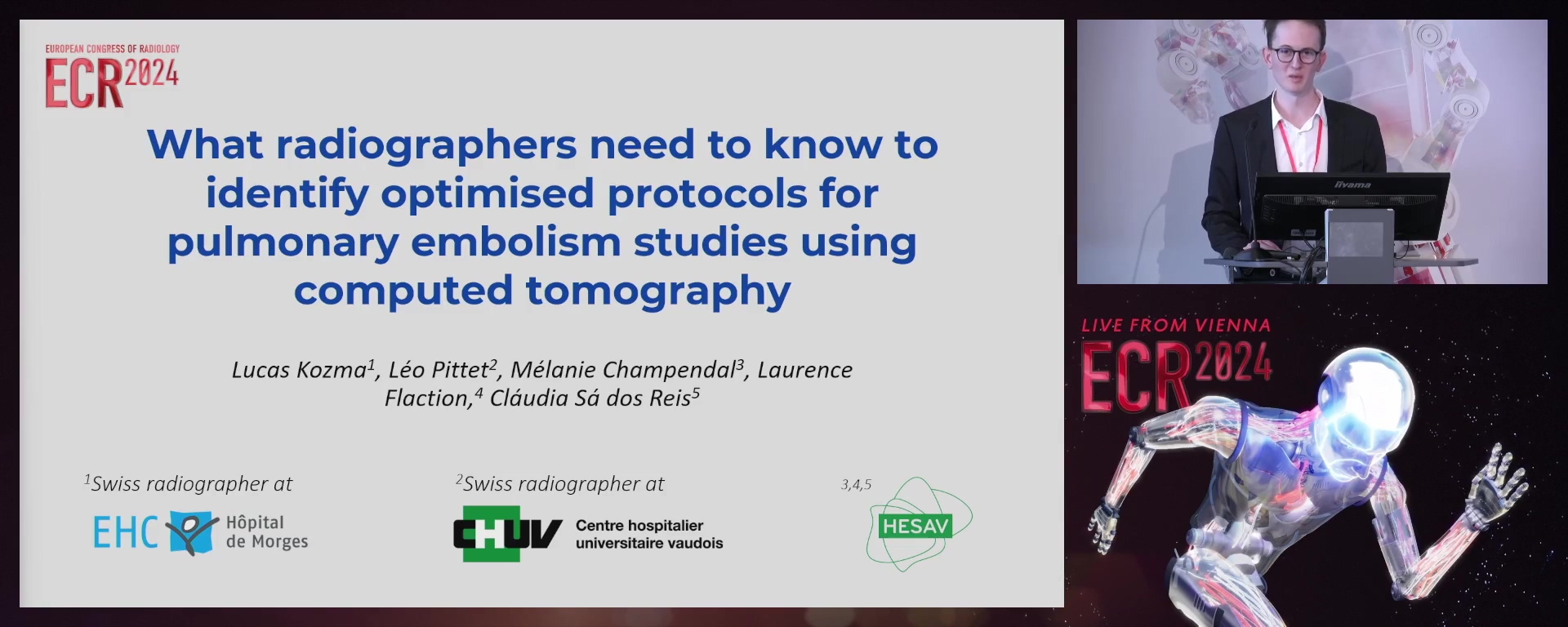
What radiographers need to know to identify optimised protocols for pulmonary embolism studies using computed tomography
Lucas Kozma, La Croix sur Lutry / Switzerland
Author Block: L. Kozma, L. Pittet, M. Champendal, L. Flaction, C. S. D. Reis; Lausanne/CH
Purpose: CTPA is the preferred method for assessing pulmonary embolism, but protocol variations result in huge radiation doses variations. This study synthesised optimised CTPA protocols to improve radiation protection, contrast media use, and image quality identified in the literature.
Methods or Background: Optimised CTPA protocols for adult patients were used, including special groups (pregnant women; high-BMI individuals), with a focus on improving radiation protection; use of iodinated contrast agents; image quality, exam success rate were identified following JBI methods. "Embase" and "PubMed" databases were searched. Two independent reviewers performed a blinded assessment. Articles published after 2018, with over 100 participants, meeting the above criteria were included. Data was extracted and analysed, with categorised subsets and descriptive statistics being applied.
Results or Findings: Twenty-eight articles, involving 100 to 3998 participants, revealed various optimisation techniques falling into five categories: acquisition, injection, breathing, reconstruction parameters, adaptations for specific populations. Reducing scan length with a 37-48% dose reduction, high-pitch scans, dose modulation (auto-mA and ODOM), lower kV (low-kV), auto-kVp, and dual-energy methods were proposed to lower dose and enhance patient outcomes. For pregnant women, 100 kV beams were recommended, with free breathing and an increased injection rate.
Conclusion: Optimising CTPA protocols is a complex task due to the amount of parameters that is necessary to consider. It requires focused studies on specific protocol variables to gather reliable and applicable data. Regular protocol reviews are crucial for effective optimisation, to include all technical updates. Radiographers are central to the optimisation process combining their technical expertise with patient-centred care expertise.
Limitations: No experimental studies were conducted.
Funding for this study: No funding was received for this study.
Has your study been approved by an ethics committee? Not applicable
Ethics committee - additional information: No information was provided by the submitters.
RPS 1014-12
7 min
Enhancing radiation safety for nuclear medicine radiographers: the impact of introducing the automated radiopharmaceutical preparation and administration system
Serena Pichierri, Milan / Italy
Author Block: S. Pichierri, G. R. Bonfitto, A. Roletto, L. Bombelli, E. Scaramelli, S. V. Fasulo, A. Savi, M. Olivieri, D. Catania; Milan/IT
Purpose: In Nuclear Medicine (NM), radiopharmaceutical doses have traditionally been administered in unit doses, divided and administered manually by personnel. The recent shift in many hospitals towards automated preparation and injection systems aims to minimize staff exposure to radiation. However, studies on their efficacy and potential benefits to radiographers are still limited. The aim of this preliminary study is to investigate the radiation exposure of NM staff involved in automated preparation and injection of (18)F-fluorodeoxyglucose (FDG), compared to manual administration.
Methods or Background: From April to July 2023, NM radiographers' radiation exposure was recorded with electronic dosimeters during (18)F-FDG administration with the automated preparation and injection system and compared with the exposure during manual preparation and administration.
Results or Findings: By analysing the radiation exposure recorded with the electronic dosimeter provided to the radiographers, it was found that the individual whole-body radiation exposure for radiographers for each patient averaged 4,03 μSv ± 1,2 during the use of the automated preparation and injection system and it averaged 6,93 μSv ± 1,5 during manual preparation and administration. Whole-body exposure was significantly reduced with automated administration by 41,85% compared to the value associated with manual administration (p < 0,05).
Conclusion: Based on preliminary results collected in our hospital, the use of an automated preparation and injection system could greatly reduce the radiation exposure of NM radiographers involved in the administration of (18)F-FDG. The results of this study will be used by our team to increase the use of the automated radiopharmaceutical preparation and administration system and more deeply evaluate its potential benefits in terms of reduction of radiation exposure on the whole NM staff.
Limitations: No limitations were identified.
Funding for this study: No funding was received for this study.
Has your study been approved by an ethics committee? Not applicable
Ethics committee - additional information: No information provided by the submitter.
RPS 1014-13
7 min
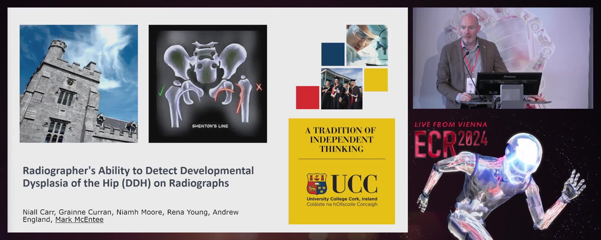
An investigation into radiographers' ability to identify developmental dysplasia of the hip (DDH) on radiographs and factors related to observer performance
Mark F. McEntee, Cork / Ireland
- Author Block: N. G. Carr1, A. England1, J. D. Thompson2, N. Moore1, R. Young1, M. F. F. McEntee1; 1Cork/IE, 2Barrow-in-Furness/UKPurpose: DDH refers to hip joint dislocation or predisposition in young children. Early detection and management are essential for effective treatment and the prevention of long-term effects. The aim of this research was to look into radiographers' abilities to detect DDH on x-ray images and identify factors that influence their performance.Methods or Background: At the European Congress of Radiology, radiographers were given a structured questionnaire which included a test bank of images. There were 25 paediatric cases in the questionnaire, and participants were asked to diagnose DDH and rate the image quality. Data were analysed to determine the sensitivity, specificity, accuracy, and area under the receiver operating characteristic (ROC) curve.Results or Findings: The image set was reviewed by 30 radiographers. The average sensitivity for diagnosing DDH was
- 2%, with a specificity of 40.2%. With an area under the curve of 0.6522, the ROC curve study demonstrated slightly better performance than chance. Collimation, lower limb position, and the absence of patient rotation were identified as key factors influencing participants' diagnostic ability. Conclusion: The study discovered that radiographers' ability to recognise developmental dysplasia of the hip (DDH) on radiographs varies and is impacted by characteristics such as training, experience, and technological consideration. Additional training and standard approaches are required to increase their diagnostic performance in DDH. DDH imaging training programmes for radiographers can improve their skills and may lead to better patient care.Limitations: Given the nature of the study, participants would have expected a prevalence of DDH within the image bank.Funding for this study: No funding was received for this study.Has your study been approved by an ethics committee? YesEthics committee - additional information: This study was approved by the Medical School SREC, University College Cork.
RPS 1014-14
7 min
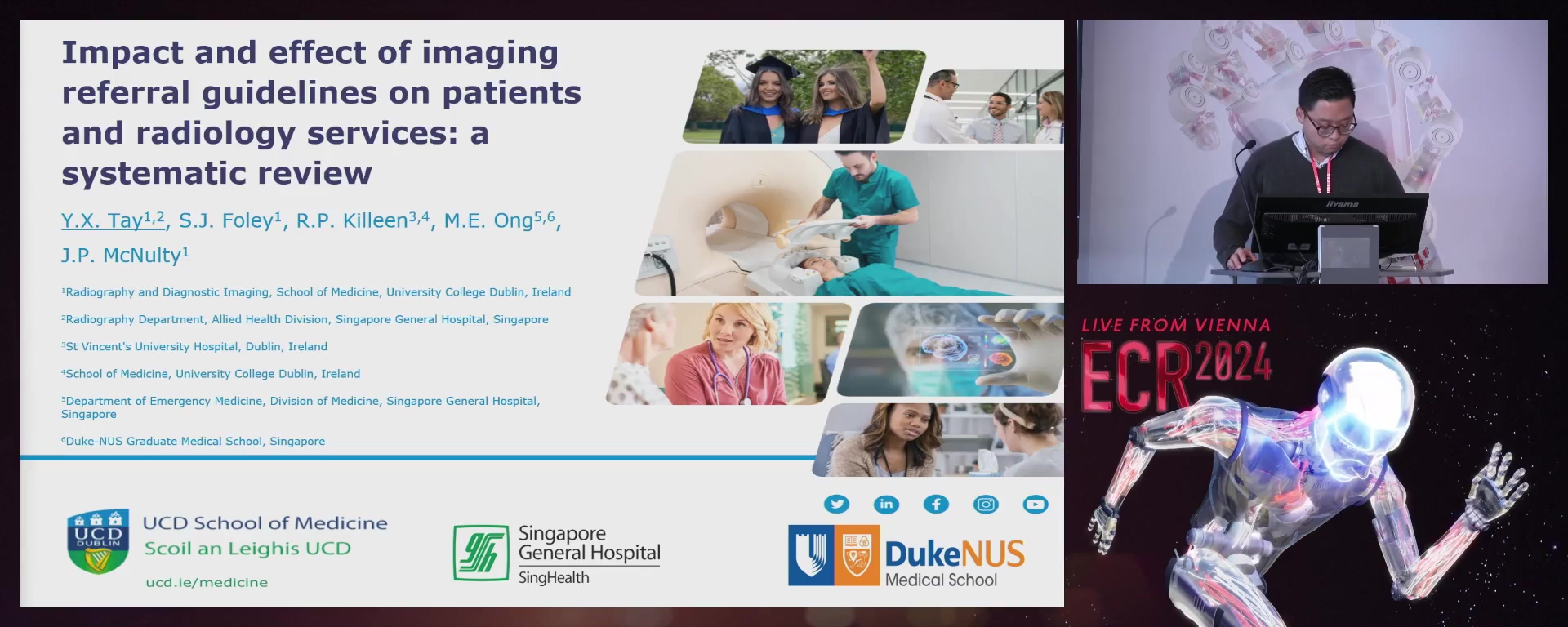
The impact and effect of imaging referral guidelines on patients and radiology services: a systematic review
Yi Xiang Tay, Singapore / Singapore
- Author Block: Y. X. Tay1, S. J. Foley2, R. P. Killeen2, M. E. Ong1, J. McNulty2; 1Singapore/SG, 2Dublin/IEPurpose: The objective of this systematic review was to offer a comprehensive overview and explore the associated outcomes from imaging referral guidelines for various key stakeholders, such as patients and radiologists.Methods or Background: An electronic database search was conducted in Medline, Embase and Web of Science to retrieve citations published between 2013 and
- The search was constructed using medical subject headings and keywords. Full-text articles and reviews written in English were included. The quality of the included papers was assessed using the mixed methods appraisal tool. A narrative synthesis was undertaken for the articles selected. Results or Findings: The search yielded 4,384 records. Following abstract, full-text screening, and removal of duplication, 31 studies of varying levels of quality were included in the final analysis. Before-and-after studies, retrospective data analysis, randomised controlled trials, interrupted time series, and cohort studies were included. Imaging referral guidelines from the American College of Radiology were most commonly used. Clinical decision support systems were the most evaluated mode of intervention, either integrated or standalone. Interventions showed reduced patient radiation doses and waiting times for imaging. There was a general reduction in radiology workload and utilisation of diagnostic imaging. Low-value imaging utilisation decreased with an increase in the appropriateness of imaging referrals and ratings and cost savings. Clinical effectiveness was maintained during the intervention period without notable adverse consequences.Conclusion: The use of evidence-based imaging referral guidelines is important to improve the quality of healthcare and outcomes while reducing healthcare costs. Imaging referral guidelines could be an essential component of improving the value of radiology in the healthcare system.Limitations: Studies included in this analysis reflect a significant degree of heterogeneity in terms of their research design, study population, interventions utilised, and outcomes evaluated.Funding for this study: Funding was received from the SingHealth Talent Development Fund and via the Singapore General Hospital Scholarship.Has your study been approved by an ethics committee? Not applicableEthics committee - additional information: Ethical review was not required as this study constitutes a systematic review of the available literature.
