Research Presentation Session: Radiographers
RPS 1714 - Enhancing patient experience and safety in medical imaging and radiotherapy
RPS 1714-3
7 min
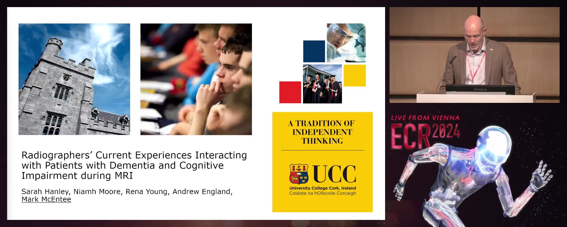
Radiographers’ current experiences interacting with patients with dementia and cognitive impairment during magnetic resonance imaging
Mark F. McEntee, Cork / Ireland
Author Block: S. Hanley, P. C. Murphy, R. Young, N. Moore, A. England, M. F. F. McEntee; Cork/IE
Purpose: Dementia is the umbrella term used to describe several conditions, causing damage to the brain. This study aims to investigate radiographers’ current experiences interacting with patients with dementia and cognitive impairment during MRI.
Methods or Background: This is a prospective quantitative study consisting of a questionnaire completed by MRI radiographers. The study location was in a single public hospital in the Republic of Ireland, performed over a six week period. The inclusion criteria required "dementia", "cognitive impairment", or "confusion" to be documented in the request form, detailed by the ward staff, or if any one of these were discovered by the MRI radiographers when the patient arrived.
Results or Findings: There were twenty questionnaires completed. The point when it was discovered the patient had dementia, cognitive impairment or confusion was predominantly through contact from the ward, accounting for 50%. Not one out of the twenty patients required someone being present during the scan. 30% of patients were agitated or distressed during the MRI examination. 15% of examinations needed to be aborted and rebooked and 25% were incomplete.
Conclusion: There were issues in discovering the patient had dementia, cognitive impairment, or confusion. There were implications regarding examinations needing to be aborted and rebooked. This is a concern as MRI is a very sought after modality. This is an area which needs to be addressed as these examination slots are vital for assisting patients along their clinical pathway.
Limitations: Identified limitation were (1) the single-centre nature of the study and (2) the relatively small sample size.
Funding for this study: No funding was received for this study.
Has your study been approved by an ethics committee? Yes
Ethics committee - additional information: This study was approved by the Medical School SREC, University College Cork.
RPS 1714-4
7 min
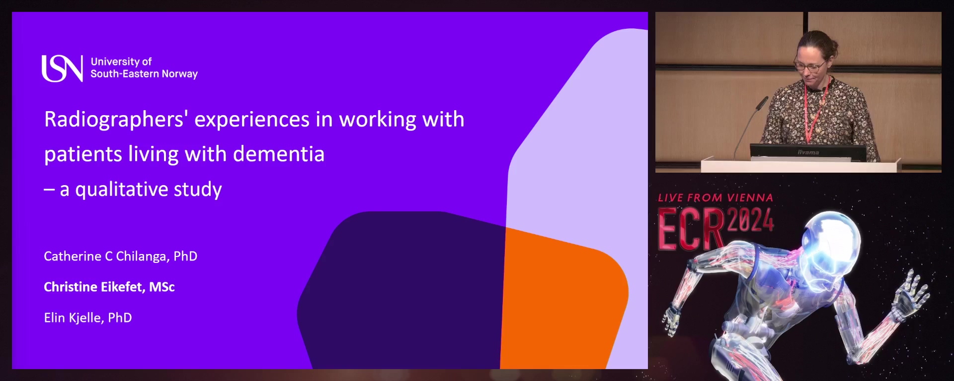
Radiographers’ experience during medical imaging of patients with dementia in Norway
Christine Eikefet, Borre / Norway
Author Block: C. C. Chilanga1, C. Eikefet2, E. Kjelle2; 1Svelvik/NO, 2Drammen/NO
Purpose: This study aimed to explore radiographers’ experiences during imaging examinations of people living with dementia in Norway.
Methods or Background: Semi-structured qualitative interviews were conducted with eight radiographers, four working in MRI or general radiography and four working in nuclear medicine, at three different hospital trusts in Norway. The interview guide included the following topics: radiographers’ experience of working with patients living with dementia, challenges faced when conducting examinations for these patients, knowledge about dementia and initiatives in the department. All interviews were verbatim transcribed and inductive content analysis as described by Elo and Kyngäs was used to analyse the data.
Results or Findings: The analysis resulted in three main categories, each with two to five subcategories. The main categories were "radiographers experience", "measures taken to accommodate for the patient" and "competencies". The radiographers frequently encountered patients with dementia in the department. The challenges they faced included a lack of information before receiving patients with dementia, communicating with the patients, and ensuring their stability during the procedure. MRI safety was of particular concern when communication and information sharing were problematic. None of the departments had any overarching procedures or training related to patients with dementia. Creating a calm environment, collaborating with carers, scheduling adequate time for examinations, and possessing good communication skills were viewed as facilitators for conducting examinations successfully.
Conclusion: Radiographers experienced managing and imaging patients living with dementia to generally be uncomplicated. The knowledge on examining patients with dementia was mostly acquired through clinical practice. However, in some cases the department’s environment, and communication problems caused stress and restlessness in patients.
Limitations: The limitation of this study is that findings from interviews with a few radiographers in Norway may not be easily generalised to other settings.
Funding for this study: No funding was received for this study.
Has your study been approved by an ethics committee? Yes
Ethics committee - additional information: The Norwegian Agency for Shared Services in Education and Research approved the treatment of personal information (project reference: 155338)..
RPS 1714-5
7 min
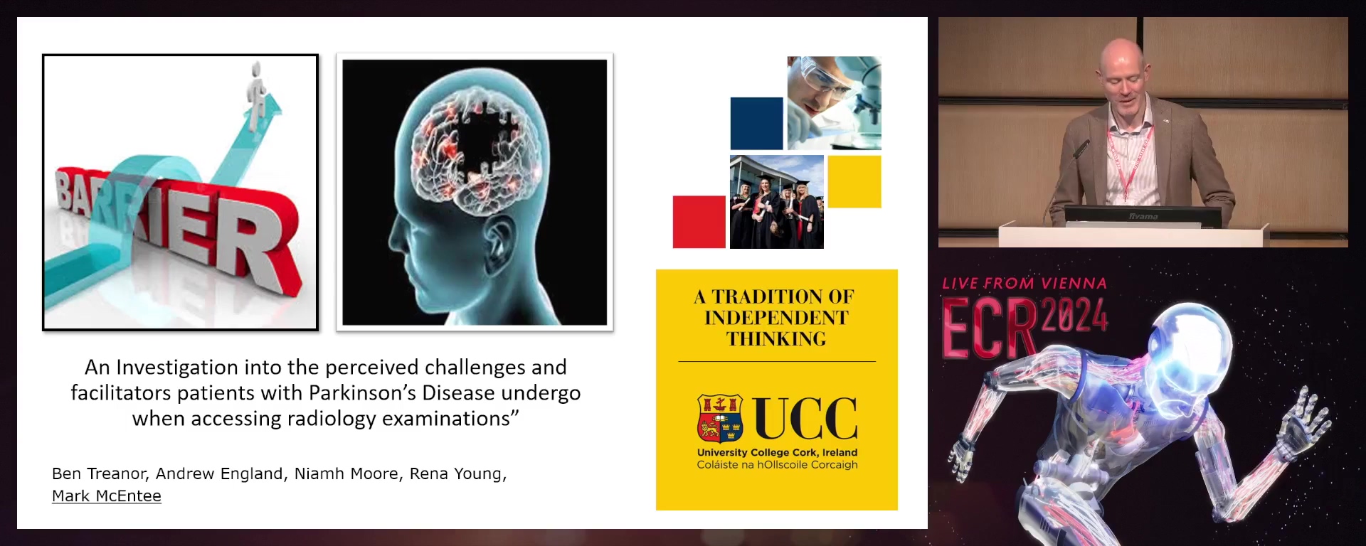
An investigation into the perceived challenges and facilitators Parkinson’s disease patients undergo when accessing radiology examinations
Mark F. McEntee, Cork / Ireland
- Author Block: B. Treanor, M. F. F. McEntee, N. Moore, R. Young, R. O'Regan, A. England; Cork/IE
Purpose: Parkinson's disease (PD) is a progressive neurological disorder characterised by a wide range of symptoms. Understanding the facilitators and barriers for patients with PD accessing radiology is paramount, as this patient group would require frequent access to imaging and the nature of their condition can pose additional challenges. The aim of this study is to investigate the perceived barriers or facilitators to radiology examinations that improve or degrade the experiences for patients with PD by conducting an experience-based focus group.
Methods or Background: This was a qualitative study in which the experiences of patients with PD were studied by conducting an audio-recorded experience-based focus group. Recruitment and data collection took place from March 2023 until April
- The primary researcher then personally validated the transcription. Following this, the primary reviewer and secondary reviewer each performed a thematic analysis of the transcription with no discrepancies found between both findings. Results or Findings: There were eight participants in this study. There were three key themes identified for the facilitators which included "supportive hospital staff", "communication" and "knowledge". There were also three main themes identified for the barriers which included "anxiety", "knowledge", and "communication". Conclusion: By addressing these facilitators and barriers, we can work towards creating a more supportive and patient-centred radiology environment for individuals living with PD. Further research is warranted to explore the experiences of patients with PD in radiology departments across different healthcare settings and geographical regions. Limitations: Responses were received from a single service user group in Ireland. Involving participants from more groups or undertaking an international study would be advantageous. Funding for this study: No funding was received for this study. Has your study been approved by an ethics committee? Yes Ethics committee - additional information: This study was approved by Medical School SREC, University College Cork.
RPS 1714-6
7 min
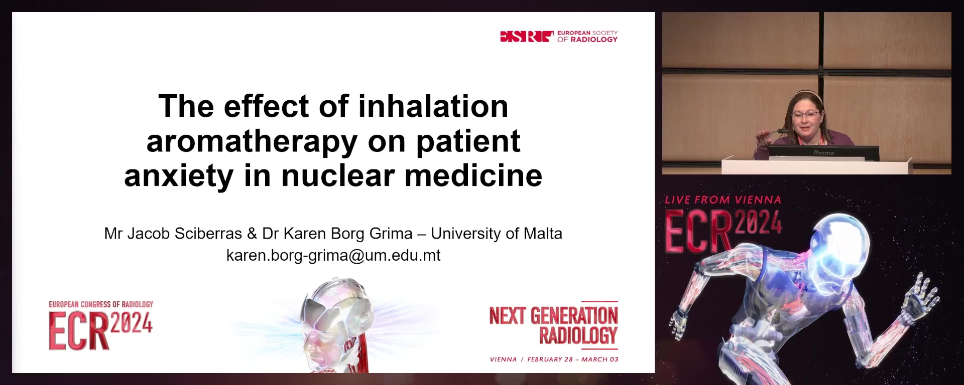
The effect of inhalation aromatherapy on patient anxiety in nuclear medicine
Karen Borg Grima, Naxxar / Malta
- Author Block: J. Sciberras, K. Borg Grima; Msida/MT
Purpose: The aim of this study was to evaluate if chamomile intervention has an impact on the anxiety levels of patients undergoing a bone or thyroid scan in nuclear medicine.
Methods or Background: A quantitative, experimental, prospective and cross-sectional design was employed for this study. Fifty participants who complied with this study’s inclusion and exclusion criteria were enlisted. Using convenient sampling, the participants were equally distributed between the experimental and control group. In both groups, routine protocol was followed when scanning the patients, apart from those in the experimental group, who were additionally introduced to the chamomile smell. Following this, the State-Trait Anxiety Inventory Questionnaire was used to measure the participants’ anxiety level before and after the scan, in both groups.
Results or Findings: Participants in both the experimental and control groups experienced higher pre-scan state anxiety (
- 84, 40.20) scores when compared to post-scan state anxiety scores (26.24, 37.12). Furthermore, the post-scan state anxiety score of the experimental group was significantly lower compared to the control group’s post-scan state anxiety score (P<0.001). In addition, when analysing the difference in anxiety levels between genders a statistical significance was noted. The results indicated that females were more vulnerable to anxiety reactions. Conclusion: From this study’s findings, there was statistical significance which indicated that chamomile intervention was effective at lowering the patients’ anxiety levels in nuclear medicine. Additionally, the results of this study suggested that chamomile intervention should be used in clinical practice to reduce patients' stress levels since it is non-invasive and cheap. Limitations: The small sample size used in this study could have influenced the accuracy of the results. For future research, a larger sample size should be applied and data should be collected over a longer period of time. Funding for this study: No funding was received for this study. Has your study been approved by an ethics committee? Yes Ethics committee - additional information: This study was approved by the University of Malta Research Ethics Committee (UREC), and by the Data Protection officer of a state general hospital where this research was conducted.
RPS 1714-7
7 min
Evaluation of the influence of music in women undergoing radiation therapy
Magda Cruz Ramos, Olhao / Portugal
- Author Block: M. C. Ramos1, A. Rosa2, F. Serra1, A. F. Abrantes2, L. P. V. Ribeiro2, S. I. Rodrigues2; 1Olhao/PT, 2Faro/PT
Purpose: Breast cancer is the most commonly diagnosed cancer among women worldwide. Patients often experience anxiety, particularly during radiotherapy, due to the disruptions in their daily routines and the associated side effects. Music therapy has emerged as a promising psychosocial intervention, offering potential benefits such as reduced anxiety, pain relief, and an improved quality of life for cancer patients. The primary aim of this research was to conduct a comprehensive examination of how these factors collectively impact anxiety levels in breast cancer patients undergoing radiotherapy.
Methods or Background: This study assume a comparative analysis between two groups of breast cancer patients. The assessment tools utilised consisted of a 'yes' system, a concise interview, the STAI (State-Trait Anxiety Inventory) questionnaire, and a visual analog comfort scale. To assess anxiety levels, the STAI questionnaire was administered twice: at the initiation of the treatment regimen and upon its completion.
Results or Findings: This study assessed anxiety levels, heart rate, education status, and comfort levels, along with examining the influence of music on anxiety during radiotherapy. The findings indicated no noteworthy age-related differences, but they did reveal a moderate correlation between anxiety and heart rate in the experimental group. No significant correlations were identified between anxiety and education status or comfort levels. Regarding the impact of music on anxiety between the groups, lower mean values of final anxiety were observed in the experimental group (
- 00 ± 2.94) compared to the control group (68.65 ± 3.69). Conclusion: While this study did not yield statistically significant differences in anxiety reduction through music during radiotherapy, its findings remain valuable and instructive. The feedback transmitted by the patients of the experimental group was shown to be favourable for the use of music, regardless of whether the results were significant or not. Limitations: No limitations were identified. Funding for this study: No funding was received for this study. Has your study been approved by an ethics committee? Yes Ethics committee - additional information: This study received authorisation from the institutional board.
RPS 1714-8
7 min
Using the e-Delphi method to develop informative nutritional leaflets for patients undergoing radiotherapy to the head and neck
Duncan Munro, Haz-Zebbug / Malta
Author Block: D. Munro, G. V. Dijk, J. G. Couto; Msida/MT
Purpose: Patients diagnosed with head and neck (HN) cancer undergoing radiotherapy (RT) frequently encounter distressing side effects that significantly impede their ability to consume food. This often results in deteriorating nutritional status and subsequent weight loss. The primary objective of this study was to evaluate the efficacy of a modified e-Delphi approach in creating informative dietary advice pamphlets aimed at addressing these side effects, while garnering consensus among healthcare professionals.
Methods or Background: An e-Delphi methodology was employed, involving six participants representing various healthcare professions specialising in HN patient care (radiographers, nurses, oncologists). These participants were tasked with providing feedback on four dietary leaflets tailored to specific symptoms, all meticulously crafted based on an extensive prior literature review. Following each round of feedback, the participants' recommended modifications were incorporated. Significant alterations to the leaflets were subjected to participant voting before implementation.
Results or Findings: After three rounds of deliberation, unanimous consensus was achieved, as all participants expressed a "highly likely" inclination to incorporate the leaflets into their clinical practice. The majority of the participants' suggestions were consistent with existing literature. The only change that deviated from the literature, and was accepted through voting, pertained to sugar consumption.
Conclusion: The participants successfully attained consensus and developed leaflets aligned with literature recommendations, which they deemed suitable for clinical application. This variation of the e-Delphi method demonstrated its efficiency in establishing consensus among healthcare professionals concerning patient information resources.
Limitations: The participant pool could have been larger. However, all the radiotherapy professions were represented in the sample. Including a dentist could have been beneficial.
Funding for this study: No funding was received for this study.
Has your study been approved by an ethics committee? Yes
Ethics committee - additional information: The researchers submitted the research for records to the University of Malta’s University Research Ethics Committee (reference number: FHS-2022-00039) following a self-assessment that showed that the research was low risk and did not require formal ethical approval.
RPS 1714-9
7 min
Radiographers’ knowledge, clinical expertise and application of pain management strategies in the radiology department
Andrew England, Cork / Ireland
Author Block: R. O'Regan, N. Moore, B. Treanor, R. Young, M. F. F. McEntee, A. England; Cork/IE
Purpose: Pain is a widely encountered medical concern that necessitates effective pain management interventions. Pain management challenges are reported within the literature as having a significant impact on medical practice, healthcare professionals, radiology departments, and the healthcare system. Radiographers frequently image patients who are in pain and thus require a heightened awareness and focus on this crucial issue. This study aims to evaluate radiographers' knowledge, clinical expertise, and challenges associated with pain management in the radiology department.
Methods or Background: A qualitative research study consisting of a single focus group was conducted with six participants. The research questions focused on pain management challenges in the radiology department, with a particular emphasis on identifying radiographers' current practices and strategies to address these obstacles. The focus group study was moderated and video-recorded to facilitate comprehensive analysis. The data obtained from the study was transcribed and thematically analysed.
Results or Findings: Data analysed from the focus group identified four main themes: consequences of pain management, communication, professional experience, and barriers. In addition, the study identified there are no current protocols, policies, and guidelines in practice for effectively managing the challenges associated with imaging patients in pain within the radiology department.
Conclusion: The focus group study revealed an array of concerns related to the challenges associated with imaging patients in pain within the radiology department. The primary consequences identified were stress experienced by both staff and patients, the potential for suboptimal images, patient safety concerns, and negative effects on image quality. The results of this research study demonstrate how patient discomfort negatively affects examinations and identifies the potential consequences for radiographers aspiring to achieve optimal gold standard imaging.
Limitations: Identified limitations were: (1) the small sample size and (2) that participants were from a single hospital group in Ireland.
Funding for this study: No funding was received for this study.
Has your study been approved by an ethics committee? Yes
Ethics committee - additional information: This study was approved by Medical School SREC, University College Cork.
RPS 1714-10
7 min
Deaf-friendly MRI: radiographers' communication toolkit
Eleonora Stefani, Susegana (TV) / Italy
Author Block: E. Stefani, M. Centenaro, G. Cappellina; Treviso/IT
Purpose: Equality is vital in healthcare and communication is part of It. Most systems are adequately prepared to communicate with certain type of patients but they don’t represent the majority of them. It is necessary to adjust communication to all types of patients. To tailor radiographers' communication to all types of patients is essential to ensure patient safety, compliance and image quality. The study aims to identify tools to improve radiographers' effective communication with deaf patients who undergo MRI examination.
Methods or Background: This was a qualitative study. One focus group has been created including radiographers from three radiology departments in a hub and spoke organisation. A total of 25 radiographers participated in the data collection. Data was analysed using ATLAS.ti software.
Results or Findings: 85% of radiographers feel a sense of discomfort when dealing with deaf patients and only 20% of them know about policies supporting deaf patients in the radiological divisions they work in.
Communication failures may occur due to: lack of knowledge about how to interact with deaf patients (90% of radiographers); difficulty in calling deaf patients in the waiting room (35%); and scheduling LIS interpreters (60%).
Three tools have been identified in support of radiographers: (1) a visual system to call patients; (2) guidelines with essential points for interacting with the deaf patient; (3)
a video in LIS concerning safety in MRI has been realised.
Conclusion: Communication is a big part of the success of MRI radiological examination. Radiographers are the main interface to patients and it is important to tailor communication approaches to their needs and to get feedback from them. Tools supporting inclusion for deaf patients optimise image quality and enhance patient experience.
Limitations: It would be worth assessing radiographers both within the public and private sectors.
Funding for this study: No funding was received for this study.
Has your study been approved by an ethics committee? Not applicable
Ethics committee - additional information: No information provided by the submitter.
This course is only accessible for ESR Premium Education Package subscribers.
Anastasia Sarchosoglou
Athens / GreeceAna Blanco Barrio
Murcia / Spain
Mark F. Mcentee
Cork / IrelandChristine Eikefet
Borre / NorwayKaren Borg Grima
Naxxar / MaltaMagda Cruz Ramos
Olhao / PortugalDuncan Munro
Haz-Zebbug / MaltaAndrew England
Cork / IrelandEleonora Stefani
Padova / Italy

