Research Presentation Session: Radiographers
RPS 314 - Innovative imaging practices and patient-centred care: radiographers at the forefront of diagnostic excellence
7 min
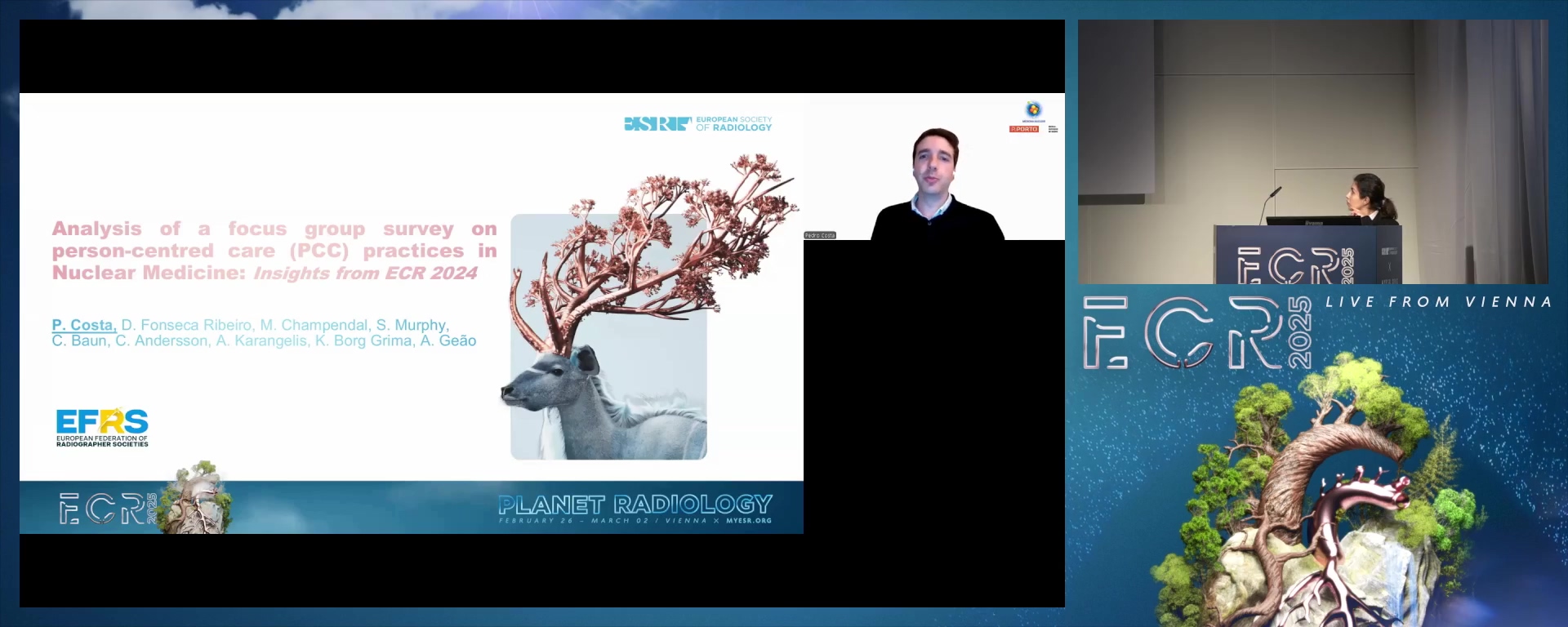
Analysis of a focus group survey on person-centred care (PCC) practices in Nuclear Medicine: insights from the European Congress of Radiology 2024
Pedro Silva Costa, Porto / Portugal
Author Block: P. Costa1, D. Fonseca Ribeiro2, M. Champendal3, S. Murphy4, C. Baun5, C. Andersson6, A. Karangelis7, K. Borg Grima8, A. Geão9; 1Porto/PT, 2London/UK, 3Lausanne/CH, 4Dublin/IE, 5Odense/DK, 6Uppsala/SE, 7Patra/GR, 8Naxxar/MT, 9Lisbon/PT
Purpose: This study aimed to gather insights into the understanding, implementation, and challenges of Person-Centered Care (PCC) practices among Nuclear Medicine professionals in Europe. Additionally, the focus group validated the questions used in the survey, serving as the ground work for a forthcoming European-wide study involving Nuclear Medicine Radiographers.
Methods or Background: A focus group survey was conducted during the European Congress of Radiology 2024. The participants included Radiographers and Nuclear Medicine Radiographers/Technologists from various European countries. The survey covered demographics, professional background, understanding of PCC, its application in clinical settings, and factors aiding or hindering its implementation. The focus group also provided some informal feedback to validate the survey questionnaire for future use.
Results or Findings: Thirty-two participants participated in this focus group and contributed to the preliminary survey, with n=5; 45% Radiographers and n=6; 55% NM Radiographers/Technologists . Most participants had between 1-10 years of experience. Participants were from Albania (9%), Italy (27%), Denmark (36%), Malta (9%), and Portugal (3%). Reported Key factors aiding PCC implementation included good communication (45%), empathy (40%), and sensitivity to patient characteristics (36%). The reported barriers to the implementation of PCC included burnout (82%), complexity of procedures (36%), and time constraints (27%). Additionally, the focus group provided feedback on the questions used while aiding to improve the survey set-up.
Conclusion: The findings revealed a strong recognition of the importance of PCC among Nuclear Medicine professionals, but also significant challenges such as burnout and time constraints which could hinder its implementation. Recommendations included, amongst others, the use of the validated questionnaire to gain a broader understanding of PCC practices in Nuclear Medicine across a wider spectrum of European countries.
Limitations: The small sample size and the limited geographical spread of the participants.
Funding for this study: No funding was received for this study.
Has your study been approved by an ethics committee? Not applicable
Ethics committee - additional information: Not applicablw
7 min
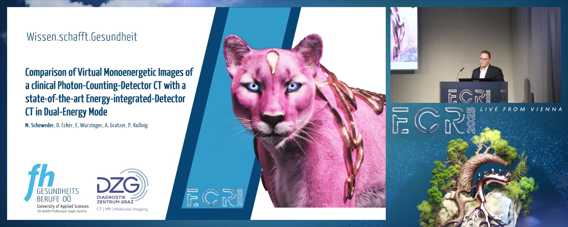
Comparison of Virtual Monoenergetic Images of a clinical Photon-Counting-Detector CT with a state-of-the-art Energy-integrated-Detector CT in Dual-Energy Mode
Mario Scheweder, Linz / Austria
Author Block: M. Scheweder1, D. Ecker1, E. Wurzinger2, A. Gratzer2, P. Kullnig2; 1Linz/AT, 2Graz/AT
Purpose: Photon-counting detector computed tomography (PCD-CT) is a promising novel technique for clinical CT, with new opportunities for image optimization while reducing radiation dose compared to conventional energy-integrated detector CT (EID-CT). Therefore, a PCD-CT and an EID-CT were compared to assess the image quality on spectral data and image reconstructions. The goal was to explore technical potentials for clinical practice.
Methods or Background: A whole-body phantom was scanned on an EID-CT in Dual-Energy (DE) Mode and a PCD-CT with similar CTDIvol. Virtual monoenergetic images (VMI) were processed at 16 keV levels (40-190 keV) and different reconstructions. Signal-to-noise Ratio (SNR) and contrast-to-noise ratio (CNR) ROIs were evaluated from liver equivalent tissue for each level and reconstruction. Mann-Whitney U test was used to compare image quality. Besides, a dose-reduced PCD-CT scan was compared to the EID-CT scan.
Results or Findings: PCD-CT-VMI data show significantly higher (all p<.05) SNR and CNR (all p<.001) than EID-CT across all keV levels and reconstruction methods. However, SNR and CNR highly depended on the reconstruction method and keV level. PCD-CT SNR/CNR exceeded ≥80 keV (SNR: +7% to +502% / CNR: +3% to +801%). A 40% dose-reduced PCD-CT scan provided higher dose-normalized SNR against the EID-CT scan at 100-190 keV (+35% to +272%). However, at 40-90 keV, PCD-CT had lower SNR (-53% to -1%).
Conclusion: PCD-CT-VMI demonstrate higher SNR/CNR capabilities compared to EID-CT in DE-Mode. This advantage can be used to optimize scan setups regarding radiation exposure. Few studies have compared the VMI data of PCD-CT and EID-CT for this purpose. However, in addition to these promising objective results, subjective image assessment by radiologists is necessary to clarify diagnostic accuracy.
Limitations: This project is a phantom study and requires further clinical research to apply findings in practice.
Funding for this study: This project was internally funded by the University of Applied Sciences for Health Professions Upper Austria. Project-Number: P-2002-003
Has your study been approved by an ethics committee? Yes
Ethics committee - additional information: The project was evaluated by the core team of the Institutional Review Board of the University of Applied Sciences for Health Professions Upper Austria, with the following conclusion: "There are no objections to the execution of this study in its current form". IRB-Number.: A-2022-018
7 min
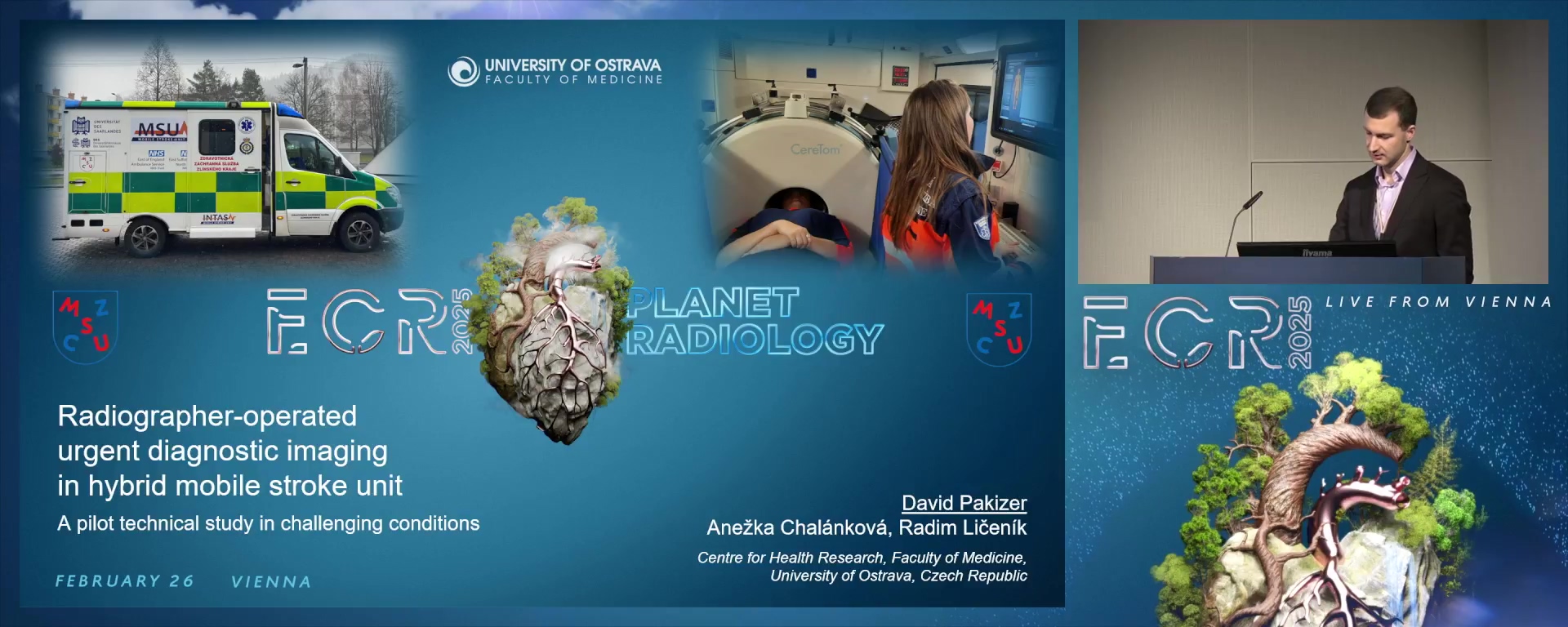
Radiographer-operated urgent diagnostic imaging in hybrid mobile stroke unit: a pilot technical study in challenging conditions
David Pakizer, Havířov / Czechia
Author Block: D. Pakizer1, A. Chalánková2, R. Líčeník3; 1Ostrava/CZ, 2Olomouc/CZ, 3Peterborough/UK
Purpose: Diagnostic assessment shift and treatment at emergency site proved beneficial for acute stroke or other neurological patients by using hybrid-mobile stroke unit (h-MSU) ambulance with mobile computed tomography (CT), portable ultrasound (US), and telemedicine onboard. We aimed to determine feasibility, safety, and efficacy of radiographer-operated h-MSU CT/US in advanced prehospital work-up for patients with acute neuroemergencies in challenging geographical/weather conditions.
Methods or Background: In our pilot prospective open-label cohort study, h-MSU was available constantly for 19 consecutive days (November/December 2023) in Czechia mountain/rural region. Patients were examined by CT intracranially in standby ambulance. Extracranial carotid US underwent selected stable patients with time of transport >30min to stroke center (ambulance on move). The h-MSU CT/US efficacy was compared with standard in-hospital modalities; feasibility and safety were assessed.
Results or Findings: Of 46 patients, 37 brain CT, 1 intracranial CT angiography, and 8 extracranial carotid US examinations were conducted. CT helped find contraindications for thrombolysis in 3 patients; 6 patients received the treatment. Of 106 CT scans, only 3% of full examinations had to be repeated. Mobile-CT mean radiation dose was only slightly higher compared to in-hospital CT, findings and image quality were similar. Low temperatures and uneven mountainous terrain were responsible for 4% of repeat CT scans. Good-quality US images were achieved and no hemodynamically significant stenosis was found but approximately 15min extracranial carotid examination was needed. Moreover, 2 US-guided cannulations were performed.
Conclusion: Mobile-CT and portable US onboard of h-MSU were safe, feasible, and effective modalities for neuroemergency patients and proved to be beneficial for time reduction and faster treatment decision-making in challenging geographical/weather conditions.
Limitations: Older age of mobile-CT/US, CT contrast agent only for study second-half, low patient number examined by US, and lack of Doppler US.
Funding for this study: None.
Has your study been approved by an ethics committee? Yes
Ethics committee - additional information: Tomas Bata Hospital Zlin Ethics Committee (approval 2023-66)
7 min
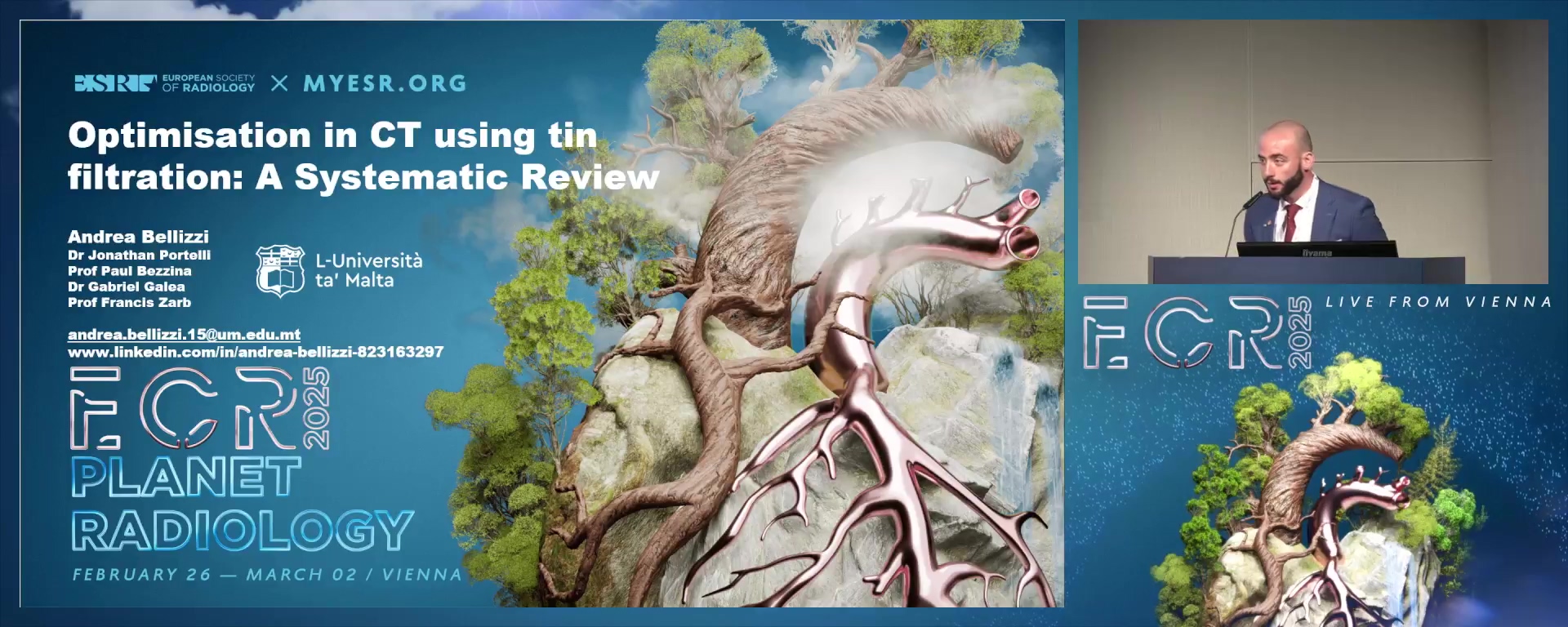
Optimisation in CT using tin filtration: A systematic Review
Andrea Bellizzi, Mosta / Malta
Author Block: A. Bellizzi, J. L. Portelli, P. Bezzina, G. Galea, F. Zarb; Msida/MT
Purpose: To identify which non-contrast CT examinations benefit from image quality and radiation dose optimisation using tin filtration (TF) and which optimisation strategy is best suited for this purpose.
Methods or Background: The review was registered in PROSPERO, and used PICO to create the research question, and exclusion/inclusion criteria. From the research question, MeSH search terms were obtained and inputted into five electronic databases: PUBMED, Scopus, CINAHL complete, Cochrane Central Register of Controlled Trials and Health & Medical Collection. Studies identified from the search were loaded into Covidence and reviewed by a team of 3 experts using PRISMA guidelines. The Joanna Briggs Institute (JBI) critical appraisal tool was used to evaluate the studies while data extraction was performed using a self-designed validated tool.
Results or Findings: From the retrieved 1479 studies, 410 were found to be duplicates leaving 1069 studies for title/abstract screening. Subsequently, 130 studies were included for full text-review, with a final 84 studies included for evaluation. TF was used to optimise scanning in 14 protocols. Scan parameters used in conjunction with TF as an optimisation strategy were: iterative reconstruction (IR) level, tube voltage (kV), pitch, rotation time and reference mAs. Use of TF achieved a significant dose reduction ranging from 17-95% in all protocols.
Conclusion: TF is an efficient dose reduction technique in non-contrast CT examinations, but has limitations meriting consideration in terms of objective image quality. These limitations could potentially be solved by varying the IR level, however more studies are needed for this to be confirmed.
Limitations: Six studies could not be retrieved. A meta-analysis could not be conducted due to the heterogeneity of the studies. Paediatrics and CT protocols using IVCM were excluded.
Funding for this study: No funding was received for this study.
Has your study been approved by an ethics committee? Not applicable
Ethics committee - additional information: None required - systematic review.
7 min
The use of Ultra-Low Dose Computed Tomography in the diagnosis of Suspected Physical Abuse in paediatric patients - A phantom study
Niamh Moore, Cork / Ireland
Author Block: E. K. Mahon, A. A. Mohammed, A. England, R. Young, N. Moore, G. A. Curran, M. F. Mcentee; Cork/IE
Purpose: Suspected physical abuse (SPA) poses a global threat to children, particularly those under the age of two. Current practices utilise conventional radiography skeletal surveys (SSs) to help with the diagnosis and management of SPA. The recent exploration of ultra-low dose CT (ULDCT) could improve diagnostic accuracy of SPA whilst minimising radiation exposure and thus is the focus of this study.
Methods or Background: CT datasets were acquired on a paediatric phantom using ULDCT (DLP=1.49 mGycm2) and standard-dose (DLP=22.92 mGycm2) CT protocols. Participants (radiographers and radiologists) subjectively scored the image quality (IQ) of both protocols using a 5-point Likert scale. Mann-Whitney U tests assessed significant differences in IQ between protocols. Participants also estimated radiation dose differences and confidence in diagnosing SPA using the CT datasets provided.
Results or Findings: Responses from 46 participants were included. Data were categorised into four anatomical areas; head/neck, thorax, abdomen/pelvis and extremities. IQ scores were consistently higher for STD when compared to ULDCT (H&N 2.7 vs 2.1; Thorax 2.8 vs. 2.2; Abdo/Pelvis 2.8 vs. 2.0 and Extremities 2.9 vs. 2.2; p<0.05). Importantly, the ULDCT protocol scored highest in “Borderline acceptable” for all regions. Interestingly, 45 (97.8%) participants underestimated the dose difference (1.49 versus 22.92 mGycm2) between the two protocols. The ULDCT protocol scored a total of 38% (88/230) confidence for the diagnosis of SPA, whereas the STD protocol scored a total of 70% (161/230) confidence.
Conclusion: Despite the IQ difference between protocols, ULDCT results are promising. Results may suggest that with adjustments to the ULD protocol 'optimisation' CT could contribute further SPA diagnosis but further research, including clinical studies, are needed.
Limitations: Phantom-based study.
Funding for this study: None.
Has your study been approved by an ethics committee? Not applicable
Ethics committee - additional information: Medical School Social Research Ethics Committee - University College Cork
7 min
Deep-learning based image reconstruction in body CT imaging: What is the real gain? A quantitative study
David Delarbre, Thônex / Switzerland
Author Block: D. Delarbre1, E. Maturana1, M. Scheffler1, J. L. Navarro Quirante1, D. Racine2; 1Geneva/CH, 2Lausanne/CH
Purpose: Compare the performance between deep learning-based Advanced intelligent Clear-IQ Engine (AICE), iterative Adaptive Iterative Dose Reduction (AIDR-3D) and filtered back projection (FBP) computed tomography (CT) image reconstruction algorithms in terms of image texture, low-contrast lesion detectability, and dose reduction potential.
Methods or Background: An abdominal anthropomorphic phantom was scanned at five computed tomography dose index (CTDIvol) level settings: 10.3, 6.4, 3.3, 2.4, and 1.9 mGy. Images were reconstructed using AICE, kernel “Body Sharp”, 1mm slice thickness, then AIDR-3D, kernel “FC08”, 1mm and 2mm slice thicknesses, and classic FBP including quantum denoising system (QDS+) reconstruction. Noise and contrast-dependent spatial resolution were assessed through noise power spectra (NPS) and target transfer functions (TTF). Texture similarity of these algorithms was evaluated using peak frequency difference (PFD) and root mean square deviation (RMSD). Lesion detectability was quantified using a non-prewhitening (NPW) observer model with eye filter. The area under the curve (AUC) and receiver operating characteristic (ROC) served as the figure of merit (FOM). Dose reduction potential for AICE 1 mm, compared to AIDR-3D with 2mm slice thickness, was calculated to achieve equivalent AUC values.
Results or Findings: At higher dose levels, AIDR-3D better preserved FBP-like noise texture. At lower doses, this difference diminished. The PFD for AIDR-3D ranged from -0.05 to -0.14, while for AICE it ranged from -0.13 to -0.17. RMSD values followed a similar trend. AICE consistently achieved higher AUC values than AIDR-3D with 1mm slice thickness, with an increasing difference as dose decreased. AICE especially demonstrated a radiation dose reduction potential of up to 45% compared to AIDR-3D with 2mm slice thickness.
Conclusion: AICE provides equivalent low-contrast lesion detectability at significantly reduced radiation doses compared to AIDR-3D, without adversely affecting noise texture.
Limitations: Constant kV, phantom morpho-type M
Funding for this study: 0
Has your study been approved by an ethics committee? Not applicable
Ethics committee - additional information: None
7 min
The effect of simulated reduced temporal resolution and motion artefacts on CT-derived cardiac left ventricular ejection fraction
Martin Weber Kusk, Esbjerg / Denmark
Author Block: M. W. Kusk1, O. Gerke2, S. Hess2, S. J. Foley3; 1Esbjerg/DK, 2Odense C/DK, 3Dublin/IE
Purpose: To test whether mid-range CT-scanners, with low temporal resolution (TR) can reliably measure left ventricular ejection fraction, (LVEF), compared to high-end cardiac scanners.
Methods or Background: 77 low-dose functional CT datasets, from a 3rd generation DSCT scanner, with 66 msec (TR) , reconstructed at 5% intervals of the entire cardiac cycle were used Cardiac MRI served as gold standard for classifying patients with potential heart failure (LVEF below 50%).
Reduced TR dataset were simulated by temporal averaging between adjacent phases, using a MATLAB script. Furthermore, in 25 artefact-free datasets, we inserted simulated discontinuity artefacts of varying magnitude and location. LVEF was measured on a clinical workstation using standard (ST) and Blood Volume (BV) modes. Absolute LVEF was compared between original and simulated images with Bland-Altman plots and t-tests, while correlation between effective TR was examined wiht Spearman rank-correlation.
Results or Findings: For BV-mode, LVEF was not significantly different between original and reduced TR images, (p=0.88) but significantly lower in ST-mode by 2.8% (p<0.01.) However, no patients were reclassified according to the 50% threshold. There was significant negative correlation between LVEF and effective TR in the ST, but not in BV mode. Motion artefacts increased the widths of 95% limits of agreement
to 5.6% in ST mode vs 2.8% in BV mode
Conclusion: Halving effective TR did not affect LVEF in the BV-mode . This mode also provided lower dispersion in the presence of motion artafacts, making it the recommended measurement method. Mid-range scanners appear suitable for LVEF measurement, , e.g. in conjunction with standard Chest-Abdomen-Pelvis scans.
Limitations: The main limitation is the lack of a true reference standard with superior temporal, and similar spatial resolution, thus making the effect of the two factors hard to disentangle.
Funding for this study: The study was funded by: The Esbjerg Fund, Research Fund of Danish Radiographers Association, Karola Jørgensen Fund
Has your study been approved by an ethics committee? Yes
Ethics committee - additional information: Regional
7 min
Patient-tailored contrast optimization in coronary CT angiography on Photon Counting CT
Tim Busselot, Leuven / Belgium
Author Block: T. Busselot, P. Giordano, W. Coudyzer, H. Bosmans, S. Dymarkowski; Leuven/BE
Purpose: Standardized injection protocols are often integrated in coronary CTA (cCTA), yet failing to account for patient variability. This study aims to introduce an evidence based injection protocol, achieving a target enhancement (expressed in terms of HU) in the overall patient population.
Methods or Background: In a first retrospective study, 162 cCTA patients, scanned on a photon-counting CT, were included. Injection parameters, demographic data and coronary enhancement were retrospectively collected and HU enhancement was measured in the aorta ascendens and proximal RCA using circular ROIs. Using the principle that iodine concentrations and HU correlate linearly, ideal iodine delivery rates (IDR) that would have provided a HU target of 500 in 55keV mono-energetic images were calculated from the HU of the original scans. Linear regression analysis was performed with the different demographic parameters and their ideal IDR, to obtain an evidence based IDR.
Secondly, 62 patients were prospectively recruited in an IRB-approved study and injected with newly proposed IDRs, wherefrom volume was calculated. To obtain lowest volumes iodine-saline dilution ratios were used. A minimal total volume of 60ml and fixed injection duration of 17s was defined to ensure complete coronary enhancement. Coronary enhancement was measured in the same ROIs as retrospectively.
Results or Findings: Ideal IDRs correlated best with bodyweight adjusted for Deurenberg’s fat formula (Pearson r=0.69), with IDR=0.0116x+0.4556
Mean coronary enhancement was 530±99HU (median: 539 and IQR:462 – 599), agreeing with the target HU of 500. Mean injected iodine volume was 51ml (median:51ml and IQR:45–57ml). All low iodine volumes could be administered under the same duration and injection rates (from 3.5–3.8ml/s).
Conclusion: A new method for injection protocol optimization, based on linear regression and integrating iodine-saline dilutions, was introduced and achieved the preset target HU enhancement.
Limitations: Mono-centric study.
Funding for this study: AI-POD project.
HORIZON Action Grant Budget-Based. Grant number 101080302.
Has your study been approved by an ethics committee? Yes
Ethics committee - additional information: UZ/KU Leuven ethics, internal number S58042
